
GLP-1 drugs can lower systemic inflammation, but direct effects on fat tissue remain unproven. In one trial, CRP fell about 30% with oral semaglutide, yet researchers did not sample fat tissue. Lower blood markers alone cannot show that inflammation within fat has changed.
The research points to three takeaways:
- Cell and animal studies suggest anti-inflammatory effects, but weight loss and other metabolic changes may explain them.
- Human fat-tissue findings are limited and mixed. Results differ by drug, fat depot, and outcome measured.
- Direct action needs stronger testing: paired biopsies, weight-loss-matched comparisons, and tests showing that responses depend on GLP-1 receptors.
This clinical overview separates laboratory, animal, and human findings - and treats tirzepatide separately because it acts on both GIP and GLP-1 receptors. These drugs should be prescribed for their FDA-approved uses, not for unproven effects on adipose inflammation.
GLP-1 and Adipose Inflammation: What the Evidence Shows
Adipose Dysfunction and Proposed GLP-1 Effects
When energy intake stays above the body’s needs, adipocytes enlarge. Their blood supply and surrounding extracellular matrix may not keep pace, leading to hypoxia, endoplasmic reticulum stress, and adipocyte death. Stressed adipocytes release CCL2 (MCP-1), which attracts macrophages. These immune cells can gather around dying adipocytes in crown-like structures. Whether GLP-1 drugs act directly on these processes - or change them through whole-body metabolic effects - remains an open question.
Inflammatory mediators, including TNF, IL-6, and IL-1β, can disrupt insulin signaling. Meanwhile, levels of adiponectin, which helps cells respond to insulin, may fall. Insulin then becomes less effective at suppressing fat breakdown. This releases more free fatty acids and adds to systemic insulin resistance. Excess collagen can also stiffen the extracellular matrix, limiting healthy tissue expansion.
Direct Adipose Signaling vs. Indirect Metabolic Effects
Establishing local activity requires validated receptor detection and evidence of downstream signaling. Bulk tissue RNA cannot show which cell type responds. Protein assays also face limits because antibodies may lack specificity and receptor levels are low. Stronger evidence would link validated receptor detection to ligand binding, downstream signaling, and a response that receptor inhibition blocks.
The evidence is stronger for some proposed effects than others.
| Proposed mechanism | Direct or indirect pathway | Supporting evidence | Evidence strength | Major uncertainty |
|---|---|---|---|---|
| Increased adiponectin and improved adipocyte function | Direct or indirect | Human ex vivo exendin-4 research found increased adiponectin without consistent improvement in inflammatory markers. | Limited human-tissue evidence | Whether the response occurs during clinical treatment , requiring careful patient selection criteria |
| Reduced macrophage inflammatory activity | Direct immune-cell action or indirect stress relief | Cell and animal studies report changes in macrophage markers and inflammatory signaling. | Preclinical support; limited human confirmation | Whether human adipose macrophages have functional receptors; whether effects occur independently of weight loss |
| Lower food intake, weight, and glycemia | Indirect | Clinical GLP-1RA studies establish these metabolic effects. | Established clinical effects | How much each change contributes to adipose inflammation |
| Reduced fibrosis or altered matrix remodeling | Mixed | Human ex vivo exendin-4 research found reduced elastin but no clear change in several collagen-related measures. | Limited and mixed | How matrix changes affect tissue function |
Weight matching alone may not isolate a direct adipose effect. When reading a study, check whether it also accounts for energy intake and glucose changes, uses paired adipose biopsies, and tests receptor dependence. Pair-fed animal studies help separate the effects of caloric intake, but they do not establish how these pathways work in humans.
The next section tests these pathways in cells, tissue, animals, and humans.
sbb-itb-7164bd9
Preclinical Research: Cells, Tissue, and Animals
The preclinical findings below offer clinical insights into potential mechanisms. They do not prove direct effects on adipose tissue in patients.
Cell and Ex Vivo Human-Tissue Studies
In 3T3-L1 adipocytes, exendin-4 increased adiponectin mRNA and secretion while reducing selected inflammatory adipokine transcripts. H-89 blocked the adiponectin response, pointing to PKA involvement - but not proving receptor specificity.
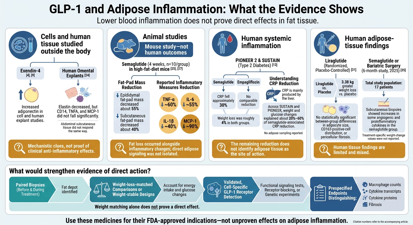
In omental explants from women undergoing elective surgery, exendin-4 increased adiponectin and reduced elastin expression and secretion. Abdominal subcutaneous tissue did not respond the same way. CD14, TNFA, and MCP-1 did not fall significantly, so the study did not show reduced inflammation. Lower elastin alone also does not establish healthier tissue remodeling.
These findings remain preclinical. Exposure may exceed levels reached in patients’ tissues, and explants lack blood flow, endocrine feedback, and immune-cell recruitment. Mixed tissue also cannot reveal which cell type responded or establish a clinical effect.
Animal models test whether these tissue signals persist when treatment acts throughout the body.
Animal Studies and the Role of Weight Loss
In male C57BL/6 mice fed a high-fat diet, 4 weeks of semaglutide treatment (n=10/group) reduced epididymal fat-pad mass by about 55% and subcutaneous fat-pad mass by about 40%.
| Model | Drug | Adipose depot | Inflammatory endpoints | Weight change | Findings | Translational limitation |
|---|---|---|---|---|---|---|
| High-fat-diet male C57BL/6 mice; 4-week treatment | Semaglutide | Epididymal and subcutaneous fat | Cytokines, macrophage infiltration, adipocyte size | Weight and adiposity decreased | Reported inflammatory measures fell approximately 60% for TNF-α, 55% for IL-6, 40% for IL-1β, and 90% for MCP-1. | Fat loss occurred alongside these changes, preventing attribution to direct adipose signaling. |
| Diet-induced-obesity mice; treatment and withdrawal | Semaglutide | Visceral and subcutaneous fat | Macrophages, crown-like structures, inflammatory genes | Weight decreased, then returned after withdrawal | Visceral inflammation declined during treatment and returned toward obese-control levels with weight regain; subcutaneous effects were limited. | Withdrawal changes both drug exposure and weight, so it cannot isolate either cause. |
| High-fat-diet/streptozotocin diabetic mice | Semaglutide | Epididymal/visceral fat | F4/80-positive macrophages and inflammatory cytokines | Reported adiposity and metabolic improvements | Reduced visceral adipose inflammation and systemic inflammatory responses were reported. | The diabetes model, dosing, and species limit generalization to human obesity. |
These models help narrow the possible mechanisms. Human biopsy data must still show whether the same tissue changes occur in people with obesity.
Testing receptor dependence requires cell-type-specific deletion, validated antagonism, or rescue experiments. Macrophage counts, cytokine proteins, and inflammatory transcripts should remain separate endpoints. Without stronger controls, preclinical models cannot separate local adipose signaling from whole-body metabolic improvement, even when their findings suggest direct action.
Human Research: Systemic Inflammation vs. Adipose Effects
Human studies test whether findings from preclinical research translate into measurable tissue changes in patients.
Clinical Trials and Adipose-Tissue Findings
Human evidence is clearer for lower systemic inflammation than for direct effects on adipose tissue. In SUSTAIN and PIONEER analyses, changes in weight and glucose explained about 20%–60% of the CRP reduction linked to semaglutide. The remaining reduction does not establish adipose tissue as the site of action.
Some trials measured only biomarkers. Others also sampled adipose tissue.
| Study design | Population | GLP-1 medicine | Adipose-specific measurements | Systemic biomarkers | Weight change |
|---|---|---|---|---|---|
| Randomized PIONEER 2 comparison | People with type 2 diabetes | Oral semaglutide versus empagliflozin | No adipose sampling reported | CRP fell approximately 30% with semaglutide, with no comparable reduction with empagliflozin. | Roughly 4% in both groups. |
| Randomized comparison with lifestyle treatment | Participants in a liraglutide-versus-lifestyle study | Liraglutide | No adipose sampling reported | Both groups had improvements in CRP and IL-1β. IL-1β reductions were comparable, but soluble CD163 fell more with liraglutide after adjustment for baseline visceral fat and triglycerides. | Similar in both groups |
| Randomized, placebo-controlled adipose study | Participants undergoing adipose assessment | Liraglutide | No statistically significant between-group differences in adipocyte size, CD163-positive-cell distribution, or pericellular fibrosis | No statistically significant differences in circulating CD163, leptin, or adiponectin | 3.38 kg (about 7.45 lb) greater loss versus placebo. |
| Six-month tissue-sampling study reported in 2025 | 17 patients undergoing semaglutide therapy or bariatric surgery. | Semaglutide | Subcutaneous biopsies showed increases in some angiogenic and proinflammatory cytokines in the semaglutide group | Metabolic markers were also assessed | No treatment-specific values were reported. |
Why Lower Biomarkers Do Not Prove Direct Adipose Action
CRP comes mainly from the liver. Lower food intake, better blood glucose control, reduced fatty-acid flux, and less visceral fat can therefore lower systemic inflammation without directly activating receptors in adipose tissue. Even when treatments produce similar weight loss, their effects on fat distribution and metabolic stress may differ.
Adjusting for weight does not prove that an effect is independent of weight loss. Weight measurements are imperfect, and weight itself is part of the treatment pathway, so adjusting for it can introduce bias. Mediation estimates also rely on assumptions about timing and unmeasured factors. Effects beyond weight loss could come from outside adipose tissue, including the brain, pancreas, liver, gut, immune system, or skeletal muscle.
When reading biopsy studies, check which fat depot was sampled, when paired samples were taken, whether the comparison group had matched weight loss, and whether endpoints were set in advance. Macrophage counts, cytokine transcripts, cytokine proteins, and fibrosis each answer different questions. Stronger evidence would pair consistent local changes with validated receptor localization and receptor-dependent signaling. Ideally, it would also show an ex vivo response blocked by GLP-1 receptor antagonism or disruption of the signaling pathway.
Conclusion: Clinical Interpretation and Research Gaps
Preclinical data suggest anti-inflammatory activity. But findings in human adipose tissue remain limited and mixed, and researchers have not established whether these effects occur independently of weight loss - even with clearer evidence of systemic improvement. Clinicians should interpret CRP as a systemic marker, not as proof of direct effects on adipose tissue.
What Clinicians Can Tell Patients
Prescribing should follow the specific product’s FDA-approved indications, established benefits, and safety requirements - not inflammatory biomarkers alone or unproven claims about adipose tissue effects.
PeptidePrescriber offers licensed healthcare professionals educational monographs, dosing protocols, regulatory references, and clinical tools. These resources are not primary research evidence, a substitute for FDA labeling, or proof of direct adipose efficacy.
Research Needed to Test Direct Adipose Effects
Testing direct effects on adipose tissue requires better biopsy-based study designs. Studies need serial biopsies before and during treatment, ideally from matched subcutaneous and visceral fat depots when feasible. Weight-loss-matched controls or weight-stable designs would help separate drug exposure from the effects of reduced calorie intake and weight loss.
Receptor detection must also be paired with functional testing using validated assays, cell-specific localization, and receptor-blocking or genetic experiments. Prespecified, standardized inflammatory endpoints would make results easier to compare across studies.
FAQs
Can a normal CRP level rule out fat-tissue inflammation?
No. A normal C-reactive protein (CRP) level does not rule out fat-tissue inflammation. CRP measures inflammation throughout the body. It cannot pinpoint inflammation in adipose (fat) tissue or confirm that none is present.
Infections, chronic inflammatory diseases, and heart, blood vessel, or kidney conditions can affect CRP levels. Results should be read alongside the patient’s weight, metabolic markers, and other possible sources of inflammation.
Should inflammation markers guide my GLP-1 treatment?
No. Inflammation markers like hs-CRP should not guide GLP-1 treatment on their own or be the main reason to change therapy. Current evidence suggests that drops in hs-CRP largely reflect weight loss - not a proven, independent anti-inflammatory effect.
Because hs-CRP is nonspecific, track it alongside weight, blood sugar, blood pressure, and overall health. Use it to monitor progress, not as a target for dose adjustments.
Does fat-tissue inflammation return after stopping GLP-1 therapy?
Research has not established whether fat-tissue inflammation returns after stopping GLP-1 receptor agonist therapy. Weight regain is common after treatment ends, but human data on inflammation after stopping treatment remain limited.
GLP-1 therapies help reduce weight and inflammatory markers such as hs-CRP. But current data do not prove that they reduce fat-tissue inflammation independently of weight loss.