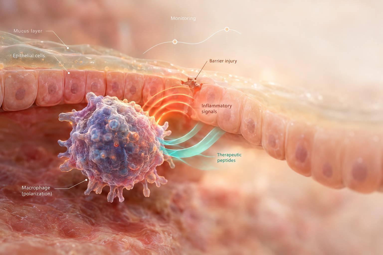
My starting point: assess gut injury - not an “M1” or “M2” label - before considering investigational peptides. Macrophages can help control microbes and repair tissue, but prolonged activation can damage the gut barrier. No validated bedside test measures their polarization.
In this guide, I focus on three questions:
- What drives injury? Excess cytokines, barrier leaks, altered gut microbes, and repair that can lead to fibrosis.
- What do peptides show? Ac2-26, gastrokine-1–related peptides, food-derived peptides, and peptide delivery systems have mostly cell and animal findings - not established human IBD outcomes. One review included 17 studies, most preclinical.
- What should you track? Symptoms, stool and blood markers, and endoscopy or imaging when indicated. Feeling better does not prove the gut lining has healed.
I also explain safety monitoring and U.S. prescribing limits: compounded drugs are not FDA-approved, and research-use-only products must not be used clinically. <u>Heavy bleeding, severe abdominal pain, dehydration, or suspected obstruction require urgent evaluation.</u>
How Macrophage Activation Sustains Gut Inflammation
Macrophage polarization in inflammatory bowel disease
2. How Macrophage Imbalance Damages the Gut
When macrophage activation persists, a signaling imbalance can turn into tissue injury.
Cytokine Excess and Barrier Damage
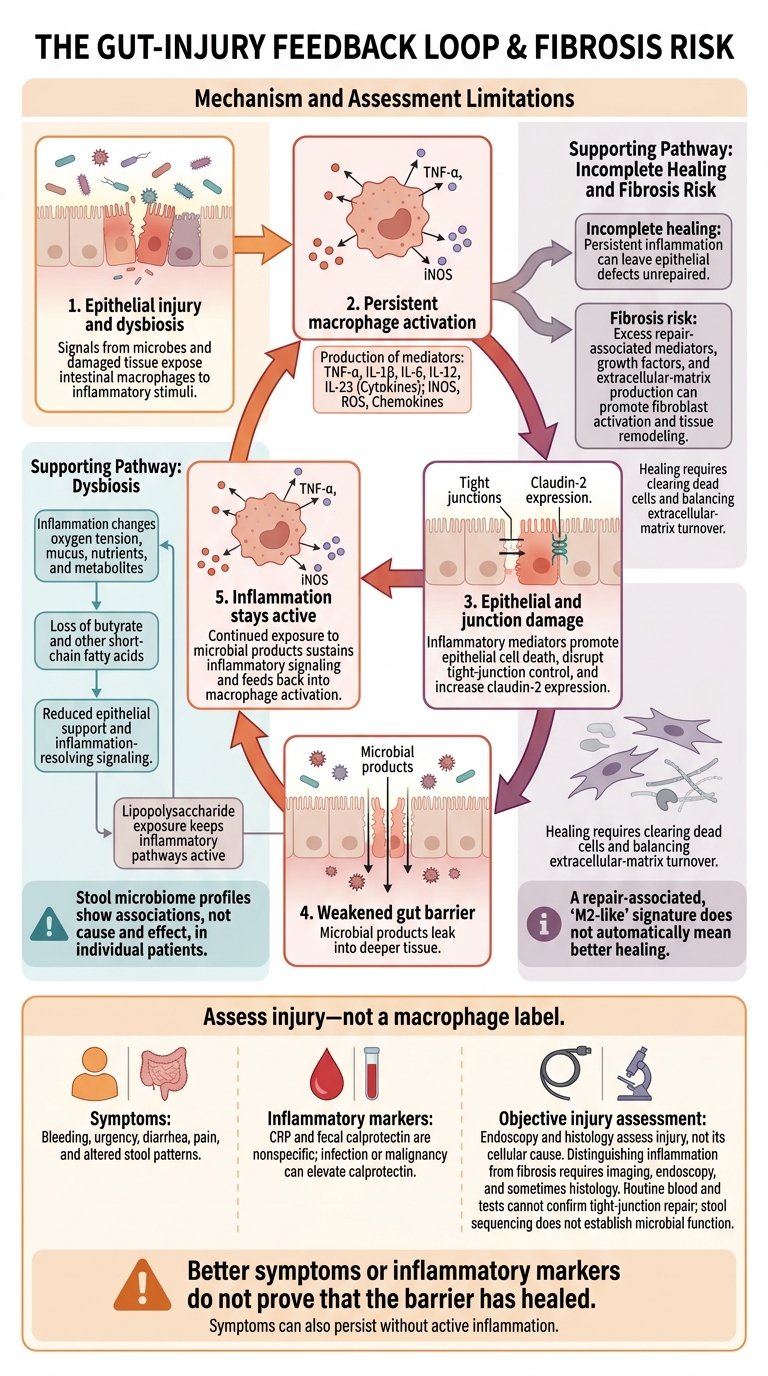
Epithelial injury and dysbiosis expose intestinal macrophages to signals from microbes and damaged tissue. These signals drive production of TNF-α, IL-1β, IL-6, IL-12, IL-23, iNOS, ROS, and chemokines that recruit leukocytes. These mediators promote epithelial cell death, disrupt tight-junction control, and increase claudin-2 expression, weakening the gut barrier. Microbial products can then leak into deeper tissue and keep inflammation active.
For peptide candidates, lower cytokine levels aren't enough. The question is whether they also reduce the tissue damage that follows.
Better symptoms or inflammatory markers do not prove that the barrier has healed. Diarrhea or pain may ease while microscopic injury remains. Symptoms can also persist without active inflammation. Assess symptoms alongside fecal calprotectin, CRP, and, when indicated, endoscopy or histology to check for healing - not just symptom relief.
Dysbiosis, Incomplete Healing, and Fibrosis
Inflammation changes oxygen tension, mucus, nutrients, and metabolites in the gut. Loss of butyrate and other short-chain fatty acids can reduce epithelial support and signaling that helps resolve inflammation. Meanwhile, exposure to lipopolysaccharide keeps inflammatory pathways active.
Stool microbiome profiles show associations, not cause and effect, in individual patients.
Healing requires clearing dead cells and balancing extracellular-matrix turnover. Persistent inflammation can leave epithelial defects unrepaired. At the other extreme, excess repair-associated mediators, growth factors, and extracellular-matrix production can promote fibroblast activation and tissue remodeling. A repair-associated, “M2-like” signature does not automatically mean better healing. Repair is the goal, but persistent inflammation can push that process toward fibrosis.
Clinical Findings and Biomarker Limits
Use these findings to assess downstream injury, not to diagnose macrophage imbalance.
| Process | Macrophage-related mechanism | Possible clinical findings | Biomarker or assessment limits |
|---|---|---|---|
| Mucosal injury | Inflammatory mediators promote epithelial cell death. | Bleeding, urgency, ulcers, histologic inflammation | Endoscopy and histology assess injury, not its cellular cause. |
| Barrier dysfunction | Cytokines disrupt epithelial survival and tight-junction control. | Diarrhea, recurrent inflammation | Routine blood and stool tests cannot confirm tight-junction repair. |
| Dysbiosis | Reduced short-chain fatty acids and increased exposure to microbial products sustain inflammation. | Bloating, altered stool patterns | Stool sequencing does not establish microbial function. |
| Cytokine excess | Inflammatory cytokine release persists. | Fever, fatigue, anemia, elevated inflammatory markers | CRP and fecal calprotectin are nonspecific; infection or malignancy can elevate calprotectin. |
| Impaired healing or fibrosis | Matrix turnover becomes dysregulated, and fibroblasts become activated. | Persistent ulcers, strictures, obstructive symptoms | Distinguishing inflammation from fibrosis requires imaging, endoscopy, and sometimes histology. |
3. Peptide Mechanisms and Evidence Gaps
The next question is whether any peptide can reduce injury without weakening host defense.
Peptide Effects on Inflammation and Resolution
Ac2-26 acts through FPR2/ALX-linked pro-resolution signaling. Cell and animal studies report reduced inflammatory signaling, IL-10-associated responses, and efferocytosis - the clearance of dead cells. These findings suggest macrophage behavior linked to inflammation resolution, but they do not prove lasting repolarization. Lower cytokine levels may also result from effects on other cell types. Lower inflammation does not prove macrophage repolarization.
In a 2024 mouse study, oral Eudragit-SBA-15-Ac2-26 at 200 micrograms once daily for 4 days reduced inflammatory-cell influx, TNF-α, and CXCL-1. It also increased IL-10 and CD163-positive macrophages and improved epithelial recovery. These results support a pro-resolution hypothesis - not established efficacy in human IBD or a dosing recommendation for humans.
Other peptides act more on epithelial recovery than directly on macrophages.
Peptide Effects on Epithelial Repair and Microbes
AMP-18/gastrokine-1 research focuses mainly on the barrier, not macrophages. Preclinical studies found preserved or increased occludin and ZO-1 in cultured human colonic epithelial cells. Mice given AMP-derived peptides also showed reduced permeability and mucosal injury. These findings do not demonstrate clinical benefit in humans.
By strengthening junctional proteins, these peptides may reduce the barrier leak that sustains macrophage activation. Full-length gastrokine-1 and its fragments need separate assessment; researchers cannot assume they produce the same exposure or effects.
Selected food-derived peptides, including buffalo-milk-derived preparations, have shown tight-junction and NF-κB effects in cell and animal research. Barrier, mucus, and antimicrobial effects are separate endpoints. Microbiome shifts may reflect reduced inflammation or barrier improvement rather than direct microbial killing. Before attributing an outcome to a peptide, check whether the study tested an identified peptide, a mixture, or a synbiotic combination.
Peptide Candidates: Evidence and Clinical Gaps
The table separates proposed mechanisms, evidence stages, and limits on applying the findings to human care.
| Candidate or class | Proposed target | Mechanism or observed effect | Evidence stage and human gut relevance | Key translation limit |
|---|---|---|---|---|
| Ac2-26 | FPR2/ALX-associated signaling | Reduced inflammatory signaling, resolution-associated markers, and epithelial recovery | Preclinical; human IBD benefit unestablished | Human pharmacokinetics unresolved. |
| AMP-18/gastrokine-1–related peptides | Epithelial junctional proteins | Preserved occludin and ZO-1; reduced permeability and injury | Translational; human gut outcomes unestablished | Fragment and full-length effects may differ. |
| Food-derived peptides | Variable immune and tight-junction pathways | Possible barrier effects; macrophage effects often indirect or unresolved | Preclinical; human gut evidence limited | Variable sequences complicate attribution. |
| Peptide nanoparticles | Stability and targeted delivery | Peptide- and formulation-dependent anti-inflammatory or repair effects | Formulation research; human gut benefit unestablished | Carrier tolerability unresolved. |
Promising preclinical results still need testing in humans. Clinicians should look for well-designed trials in patients with confirmed IBD that establish tissue exposure, pharmacokinetics, dose response, validated outcomes - including endoscopic healing - safety, and durability. Safety findings from unrelated indications do not establish safety in active colitis.
Investigational peptides remain adjunctive research tools, not substitutes for diagnosis or guideline-directed IBD care.
sbb-itb-7164bd9
4. Clinical Assessment, Monitoring, and U.S. Requirements
Moving from mechanisms to clinical care requires a confirmed diagnosis, objective monitoring, and compliance with U.S. regulations.
Baseline Assessment and Urgent Warning Signs
Establish the diagnosis before attributing symptoms to macrophage imbalance. Because this imbalance presents as injury, barrier failure, dysbiosis, and excess cytokines, assessment should focus on those effects.
Document symptom onset, stool frequency, bleeding, urgency, nocturnal stools, pain, weight change, intake, hydration, and daily function. Review prior endoscopy, imaging, pathology, treatment response, medications, immunosuppressive exposure, recent antibiotics, infection risks, allergies, pregnancy status when relevant, and comorbidities.
Consider other causes, including infection, ischemia, medication injury, celiac disease, malignancy, and functional disorders. Baseline testing commonly includes CBC with differential, CMP, CRP, albumin, and disease-appropriate stool tests. Include fecal calprotectin when IBD is suspected, iron studies for anemia or bleeding, and Clostridioides difficile testing when indicated.
Urgent evaluation is needed for heavy bleeding, dehydration, inability to take fluids, fever with systemic illness, tachycardia or hypotension, severe or quickly worsening abdominal pain, distention with vomiting or obstipation, syncope, confusion, or sepsis. Do not explain unexplained or worsening symptoms as macrophage shifts or a healing crisis.
Monitoring Symptoms, Inflammation, and Safety
Turn proposed anti-inflammatory or barrier effects into measurable clinical targets. Record symptom burden, product, formulation, concentration, route, lot information, start date, and concurrent therapies.
Set reassessment dates based on disease activity and treatment risk. Stable IBD in symptomatic remission may allow biomarker checks every 6–12 months; active disease or safety concerns call for earlier review.
These measures track the effects of macrophage-driven injury - not macrophage state itself. They assess mucosal injury, barrier dysfunction, inflammation, and treatment safety.
| Clinical objective | Measures | Limits and reassessment triggers |
|---|---|---|
| Reduce symptom burden | Daily stool count, bleeding, urgency, nocturnal stools, pain, sleep, and daily function | Symptom relief does not establish mucosal healing. Worsening bleeding, pain, fever, or dehydration needs prompt reassessment. |
| Assess systemic inflammation | CRP in mg/L | Normal CRP does not exclude intestinal inflammation; infection can increase it. Review trends alongside symptoms. |
| Assess intestinal inflammation | Fecal calprotectin in μg/g | In selected patients with ulcerative colitis in symptomatic remission, a result below 150 μg/g can help rule out active inflammation. Elevated results or disagreement between symptoms and biomarkers may require endoscopy. |
| Detect anemia or toxicity | CBC with differential | Falling hemoglobin or new leukopenia, neutropenia, or thrombocytopenia requires evaluation for bleeding, infection, or medication effects. |
| Assess nutrition and hydration | Weight, intake, hydration, albumin in g/dL, electrolytes, kidney and liver tests | Albumin also reflects inflammation, protein loss, and liver disease. Weight loss or worsening laboratory results warrants reassessment. |
| Check safety and structural disease | Administration reactions, allergy, or infection signals; disease-specific endoscopy or imaging | Review interactions with corticosteroids, biologics, immunomodulators, and anticoagulants. Persistent symptoms, obstruction concerns, or conflicting markers require objective assessment. |
Evidence Review and Prescribing Safeguards
Review human outcomes and safety separately from mechanistic findings. Verify FDA approval status, indication, formulation, route, and federal and state compounding requirements. Confirm pharmacy authorization, sterility, concentration, storage, and beyond-use date.
Compounded drugs are not FDA-approved. They do not undergo FDA premarket review for safety, effectiveness, or manufacturing quality. Research-use-only products must not be administered clinically.
Document consent and a review date. Hold or stop treatment for serious reactions, infection concerns, disease worsening, major laboratory changes, unverifiable quality, or no objective benefit within the agreed evaluation period. Escalate care to gastroenterology for diagnostic uncertainty, persistent bleeding, rising markers, malnutrition, steroid dependence, or treatment failure.
5. Conclusion: Match Mechanisms to Clinical Evidence
The clinical question is whether treatment changes measurable gut outcomes - not whether macrophages shift in theory. Macrophage imbalance sustains excess cytokines, barrier injury, dysbiosis, and, if repair persists, fibrosis.
Peptide mechanisms remain hypothetical until human outcomes confirm a benefit. A proposed mechanism does not prove efficacy. A systematic review found 17 anti-inflammatory peptide studies, most of them preclinical. Human evidence remains limited on whether these proposed immune effects improve gut outcomes without weakening host defense.
Before any peptide trial, establish the diagnosis and identify the active clinical problem. Then review human evidence and U.S. regulatory requirements, define objective endpoints up front, and plan safety monitoring. Coordinate decisions with gastroenterology and other relevant clinicians. A proposed macrophage effect should not serve as a treatment target on its own.
Seek immediate medical evaluation for severe or fast-worsening abdominal pain, persistent vomiting, marked abdominal distension, inability to pass stool or gas, heavy rectal bleeding, black stools, fainting, confusion, severe dehydration, high fever, or signs of sepsis.
Symptom relief or lower inflammatory markers do not prove mucosal healing. Endoscopic, histologic, and antifibrotic outcomes are distinct. Match each claim to the outcome actually measured.
FAQs
Can reducing macrophage inflammation weaken my gut’s defenses?
Yes, there’s a theoretical risk. Reducing macrophage-driven inflammation is often needed, but long-term blockade of pathways like NF-kB may weaken the body’s defenses against microbes and hinder repair of the gut lining. Complete inhibition has been linked to spontaneous colitis in preclinical models.
Clinical monitoring for infection is recommended when using anti-inflammatory peptides. The aim is to reduce excessive cytokine production while preserving immune function and a healthy gut barrier.
How can I tell inflammation from gut scarring?
Symptoms alone often can’t tell active gut inflammation from scarring. Check CRP, ESR, and fecal calprotectin at baseline, then every 4–8 weeks. A fecal calprotectin level below 50 μg/g helps rule out active inflammatory bowel disease; a level above 250 μg/g calls for further investigation.
If symptoms or biomarkers worsen, use endoscopy, histology, and stool studies to confirm the underlying cause rather than increasing the dose without that assessment.
What should I ask my doctor about investigational peptides?
Ask about the product’s regulatory status: Is it FDA-approved or compounded? Ask what research supports its use as a treatment, too. Many peptides lack long-term safety data in humans, with research based mainly on animal studies or in vitro (lab) testing.
Discuss possible risks, including interactions with medications you take. Ask for a clear monitoring plan with a set timeframe, using blood tests for inflammation or symptom logs to track progress and safety.