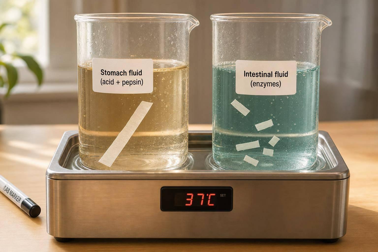
If I had to sum it up in one line: a peptide can survive the stomach and still get cut apart in the small intestine.
Here’s the short answer:
- The stomach is harsh because of low pH and pepsin
- The small intestine has more proteases and often causes more overall breakdown
- Stability in gastric fluid does not predict stability in intestinal fluid
- The main failure point helps me judge what kind of protection may fit:
- Early gastric loss: think acid stress, unfolding, or pepsin cleavage
- Later intestinal loss: think trypsin, chymotrypsin, carboxypeptidases, and brush-border enzymes
- SGF-to-SIF testing at 98.6°F (37°C) is a better first screen than testing one compartment alone
In plain terms, the stomach and small intestine damage peptides in different ways. The stomach hits first with acid plus pepsin. The intestine then adds a broader enzyme mix, often over several hours, with pH moving from about 5–6 in the duodenum to 7–8 farther along.
A few points matter most to me:
- Food changes gastric pH and residence time
- Pepsin favors sites near aromatic residues, but structure still matters
- Trypsin and chymotrypsin can cut peptides that looked stable in the stomach
- One early cut can trigger stepwise trimming
- In vitro stability is only the start; absorption can still be poor
Why oral GLP1 fail: the 1% bioavailability problem!
sbb-itb-7164bd9
Quick Comparison
| Factor | Gastric fluid | Intestinal fluid |
|---|---|---|
| Main stress | Acid + pepsin | Broader protease attack |
| Usual pH | ~1–2 fasted, higher after meals | ~5–8 depending on region |
| Main enzymes | Pepsin | Trypsin, chymotrypsin, elastase, carboxypeptidases, brush-border enzymes |
| Common failure pattern | Early unfolding and cleavage | Sequential fragmentation and trimming |
| What results may suggest | Need for enteric coating or local buffering | Need for protease protection or sequence changes |
Bottom line: I would not judge an oral peptide by stomach data alone. Where it breaks down tells me far more than whether it breaks down.
Gastric fluid: low pH, pepsin activity, and early peptide degradation
The stomach is the first big barrier for oral peptide delivery. In the fasted state, gastric pH usually sits around 1–2, which creates a harsh acidic setting that can destabilize peptides even before enzymes start cutting them apart. After a meal, gastric pH can climb into the 3–7 range and then drop again over the next 2–3 hours. That matters a lot in practice. For an oral peptide, early exposure can look very different depending on when it is taken relative to food. So the stomach is the first major survival test.
How pepsin and acid work together
Acid and pepsin work as a pair. Low pH protonates ionizable groups, which disrupts the noncovalent interactions that help hold a peptide’s structure in place. In more flexible peptides, that can lead to partial unfolding and expose internal bonds that were harder to reach before. Once that happens, pepsin has an easier target.
Pepsin tends to cleave bonds near phenylalanine, tryptophan, tyrosine, and leucine, but the outcome still depends on sequence context and structure. In plain terms, two peptides can contain similar residues and still behave very differently in the stomach. Resistance in gastric fluid depends on structure, not sequence alone. And even if a peptide gets past pepsin, that still doesn’t mean it will make it through the rest of the GI tract.
Why gastric residence time matters
Gastric residence time is not a fixed timer. It can vary by hours. Meals, especially high-fat or high-protein meals, can keep peptides in the stomach longer, which extends exposure to both acid and pepsin and increases degradation risk for compounds that might otherwise pass through more quickly in the fasted state.
The next barrier is the small intestine, where pH is higher and proteolysis becomes broader, so the pattern of failure changes.
Intestinal fluid: broader proteolysis at a higher pH
Once a peptide gets past the stomach, it enters a less acidic setting in the small intestine. But that does not make things easier. In many cases, it means the opposite.
Pancreatic bicarbonate neutralizes gastric acid, which lifts the luminal pH and switches pancreatic proteases from inactive zymogens into active enzymes. That change exposes peptides to more cleavage opportunities and a much larger mix of proteases.
Key intestinal enzymes and where they act
At the brush border, enteropeptidase converts trypsinogen into trypsin. Trypsin then activates chymotrypsinogen, proelastase, and procarboxypeptidases A and B. Proteolysis is strongest in the duodenum and upper jejunum, where pancreatic enzymes first mix with intestinal contents.
Here’s the basic split:
- Trypsin, chymotrypsin, and elastase are endopeptidases, so they cut internal peptide bonds.
- Carboxypeptidases A and B remove residues from the C-terminus.
After that first round of luminal fragmentation, brush-border aminopeptidases, carboxypeptidases, and dipeptidases keep breaking peptides down at the epithelial surface. Enterocytes can also hydrolyze absorbed fragments after uptake, which means loss from the lumen does not necessarily mean intact absorption.
Why surviving the stomach does not guarantee intestinal stability
Getting through the stomach is only part of the story. A peptide may resist pepsin and still be cut apart in the intestine once trypsin or chymotrypsin sites become exposed after gastric emptying. If lysine or arginine residues are easy to reach, the peptide may turn into a fast trypsin substrate. If aromatic residues are exposed, chymotrypsin may cleave it just as fast.
There’s also a chain-reaction effect. An early endoproteolytic cut can expose fresh cleavage sites, and then carboxypeptidases plus brush-border enzymes keep trimming the fragment step by step. That matters because intestinal exposure lasts long enough for repeated cleavage.
Small-intestinal transit lasts for hours, and one reference estimates 50% emptying at 2.5–3 hours. In experimental studies, oral peptides can disappear from intestinal fluid within minutes. That gap in exposure helps explain why stomach survival and intestinal stability often do not line up.
Gastric vs. intestinal fluid: side-by-side comparison and why degradation patterns shift
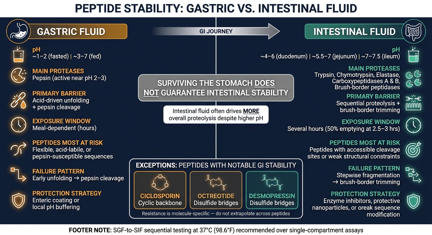
Gastric vs Intestinal Fluid: Peptide Stability Comparison
Use the comparison below to spot the main barrier first: acid, enzymes, or time in transit. The table shows which barrier tends to hit first and why degradation often starts in the stomach, then shifts to the intestine. That shift helps explain why some peptides fall apart early, while others make it through the stomach and break down later.
| Factor | Gastric fluid | Small-intestinal fluid | Clinical significance |
|---|---|---|---|
| Typical pH | ~1–2 fasted; ~3–7 fed | ~4–6 duodenum; ~5.5–7 jejunum; ~7–7.5 ileum | pH shifts alter conformation, charge, and enzyme activity |
| Main proteases | Pepsin, most active near pH 2–3 | Trypsin, chymotrypsin, elastase, carboxypeptidases, and brush-border peptidases | Intestinal broader enzyme attack is often more rapid |
| Main barrier | Acid-driven unfolding plus pepsin cleavage | Sequential proteolysis plus brush-border trimming | One barrier rarely acts alone |
| Exposure window | Meal-dependent hours | Several hours | Longer exposure increases cumulative degradation |
| Peptides most at risk | Flexible, acid-labile, or pepsin-susceptible sequences | Peptides with accessible cleavage sites or weak structural constraints | Screening in gastric fluid alone is not sufficient |
Why some peptides fail in the stomach while others fail later in the gut
The pattern is pretty straightforward. Acid loosens the peptide structure first, pepsin takes advantage of that exposed structure, and intestinal proteases keep cutting after gastric emptying. In many cases, pepsin - not acid by itself - drives much of the early gastric loss.
If a peptide gets through that first stage, it can still run into trouble later. A molecule may hold onto a compact shape at low pH or simply lack easy pepsin-sensitive sites, which lets it pass into the small intestine mostly intact. But once it gets there, the setting changes. Pancreatic enzymes switch on after gastric emptying and neutralization, and brush-border peptidases keep processing the fragments that remain.
That stepwise breakdown matters. One early cut can expose new weak points, and then the next enzymes keep trimming from there. So a peptide can survive the stomach with little damage and still break down fast in the intestine.
Six molecular traits do much of the heavy lifting here:
- acid sensitivity
- sequence
- molecular size
- flexibility
- cyclization or disulfide bridges
- compact structure
In plain terms, larger or more flexible peptides often leave more bonds open to protease attack. More compact or constrained structures can shield those bonds and slow enzymatic access. But this is molecule-specific, not a blanket rule.
Molecule-specific resistance patterns
A 17-peptide assessment of gastrointestinal stability found that most tested molecules degraded rapidly in human small-intestinal fluid, with ciclosporin, octreotide, and desmopressin as notable exceptions. Their resistance did not come from the exact same feature. Ciclosporin has a cyclic backbone, while octreotide and desmopressin contain disulfide bridges.
Those molecules are useful reference points, but they shouldn't be treated like a shortcut. The same study found that stability changed across test systems - gastric, simulated intestinal, and human intestinal fluid - and that strong performance in one setting did not reliably predict performance in another.
Testing peptide stability and applying results to oral delivery design
Once you know the likely failure point, test the peptide in the same order it would face in the GI tract: SGF first, then SIF, both at 37°C (98.6°F), with samples taken at multiple time points.
For analysis, use RP-HPLC to measure how much intact peptide is left. Then use LC-MS/MS to map cleavage sites and tell apart enzymatic breakdown from nonenzymatic loss, such as oxidation, aggregation, or adsorption to the vessel wall. Using both methods together gives a better read than relying on just one.
That said, assay setup is only part of the story. The model itself has limits. Standard SGF and SIF systems don't match fed-state pH shifts, bile, motility, mucus, or enzyme changes linked to disease. Published comparisons have also shown that SIF can degrade peptides faster than human or pig intestinal fluids, which means these results may be conservative for some peptides. So simulated-fluid data works best as a screening tool, not a stand-in for in vivo bioavailability.
What stability results mean for oral delivery strategy
The site of degradation should guide the protection plan.
- Rapid loss in SGF usually points to an acid or pepsin problem. In that case, options like an enteric coating or local pH buffering make sense.
- Good survival in SGF but rapid loss in SIF points somewhere else: pancreatic and brush-border proteases, not acid. That calls for intestinal protease protection, such as enzyme inhibitors, protective nanoparticles, or sequence modification.
There's also an easy mistake to make here. Gastric protection alone doesn't prove oral bioavailability. Even if a peptide makes it through both SGF and SIF in vitro, absorption can still be poor because of mucus, low epithelial permeability, or first-pass barriers. Stability is the starting point. It doesn't settle the whole question.
Conclusion: key differences clinicians should remember
Gastric fluid is more acidic, but intestinal fluid often drives more overall proteolysis. In other words, pH matters, but enzymes often matter more. Exenatide is a good example: it degraded when digestive enzymes were present even though it stayed stable across pH buffers.
Peptide degradation is also molecule-specific and sequence-dependent. So if one peptide fails in a certain way, that pattern shouldn't be copied over to another peptide without testing it. Sequential SGF-to-SIF studies do a better job of reflecting the two-step GI barrier than single-compartment assays, which is why they give a more useful picture early on.
FAQs
Why can a peptide survive the stomach but fail in the intestine?
Because the stomach and small intestine don’t break down peptides in the same way.
A peptide might make it through stomach acid just fine. But that doesn’t mean it will survive the next step. The stomach mainly uses pepsin to break things down, while the small intestine has a much broader mix of peptidases.
That difference matters. Once the peptide reaches the intestine, those enzymes can cleave its amino acid sequence before it’s absorbed. And if that happens, the peptide may be broken down and inactivated even though it handled the stomach’s low pH without much trouble.
How does food affect peptide stability in the GI tract?
Food can lower peptide stability by changing GI pH and triggering digestive enzymes. Gastric proteases like pepsin, along with small-intestinal brush-border peptidases, can break down many therapeutic peptides before they’re absorbed.
Some peptides, such as BPC-157, hold up better in gastric acid because of their structural properties. For oral use, clinicians often recommend taking these peptides on an empty stomach so there’s less digestive interference.
What does SGF-to-SIF testing show?
SGF-to-SIF testing simulates a peptide’s path from the stomach to the small intestine to check how well it holds up against GI breakdown.
In plain English, it shows whether the peptide stays intact after exposure to acidic gastric conditions and proteases, and then through intestinal digestion. That gives you a clearer sense of whether it can survive the GI tract or whether it may need a different delivery method to help preserve bioavailability.