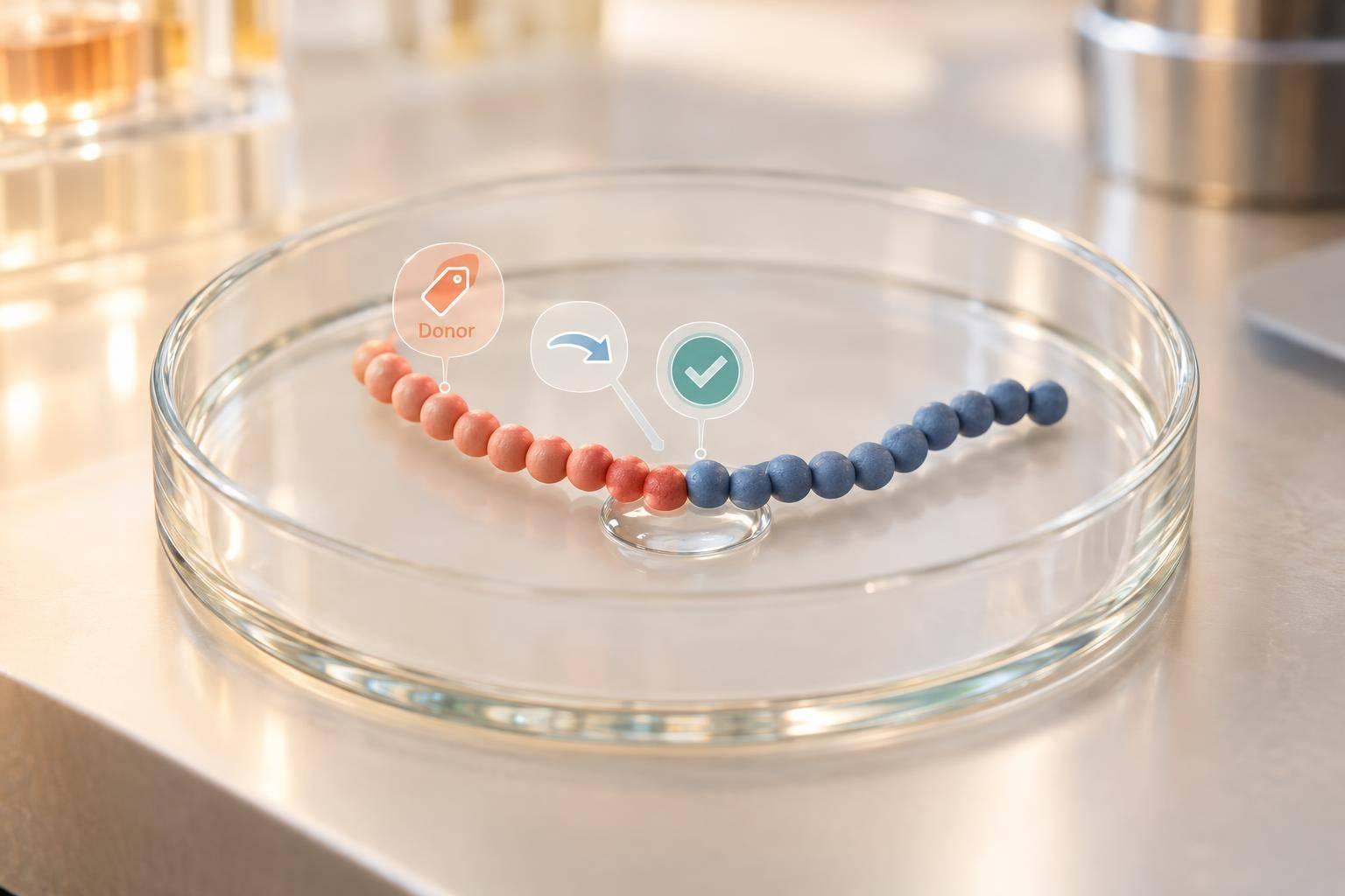
Asparaginyl ligases join peptides or close them into rings - but only when the enzyme and peptide sequences match. Start by defining the intended junction, choosing a compatible ligase, and checking that the acceptor has a free N-terminal amine. For butelase-1, the donor motif Asn–His–Val is one tested option.
A sound workflow covers 3 checks:
- Substrate fit: Match donor and acceptor sequences to the selected enzyme’s rules.
- Reaction performance: Track ligation alongside competing cleavage and unwanted products using controls and time-course samples.
- Product identity: Use chromatography, mass spectrometry, and junction mapping. Starting-material loss or the expected mass alone does not prove correct ligation.
Butelase-1, engineered ligases, and cleavage-focused AEPs differ in substrate preferences and reaction outcomes. Scaling up also requires control of enzyme supply, impurities, recovery, and batch consistency. A verified research product is not proof of safety, efficacy, or FDA approval.
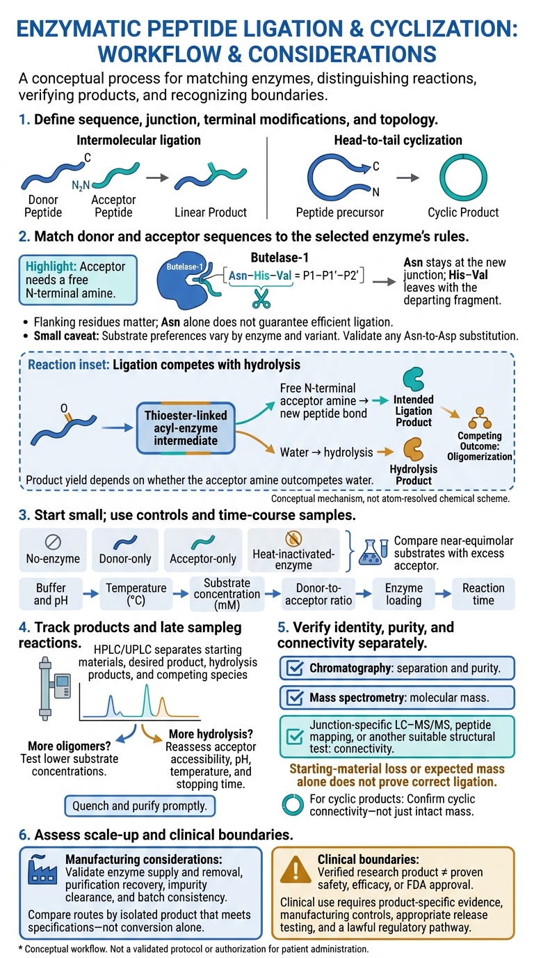
Asparaginyl Ligase Workflow: Design to Verified Product
Ligation Chemistry and Substrate Recognition
How Asparaginyl Ligases Form Peptide Bonds
Ligation depends on directing the acyl-enzyme intermediate toward peptide bond formation rather than hydrolysis. The catalytic Cys-His dyad attacks the scissile bond after a compatible P1 Asn, or P1 Asp for some enzymes under certain conditions. Cleavage releases the downstream fragment, leaving a thioester-linked acyl-enzyme intermediate.
The acceptor’s free N-terminal amine then attacks this intermediate. That step forms a new peptide bond and regenerates the enzyme. Product yield depends on whether the acceptor amine outcompetes water.
donor–Asn–X + acceptor–NH₂ → ligated donor–Asn–acceptor + X-containing leaving fragment
If water attacks the intermediate instead, hydrolysis occurs. An accessible acceptor, compatible flanking residues, and suitable reaction conditions help favor ligation.
Separate donor and acceptor peptides undergo intermolecular ligation. A single peptide with both ends available can cyclize head-to-tail. Bringing those ends close together helps cyclization, but hydrolysis and oligomerization still compete.
Butelase-1 shows how precise donor and acceptor geometry shapes this chemistry.
Butelase-1 Substrate Requirements
For butelase-1, substrate positions are written P4–P3–P2–P1 | P1′–P2′. The vertical line marks the cleavage site, and the corresponding enzyme pockets are S4–S1 and S1′–S2′.
In the donor motif Asn–His–Val, Asn sits at P1, His at P1′, and Val at P2′. Asn stays at the new junction, while His–Val leaves with the departing fragment. Flanking residues also affect recognition, so Asn alone does not guarantee efficient ligation.
Butelase-1 works in mild, near-neutral aqueous conditions. Its acceptor needs a free N-terminal amine. Acetylation, pyroglutamyl blocking, or steric shielding can reduce or redirect ligation. Follow the reported acceptor rules for the selected enzyme, and validate any Asn-to-Asp substitution.
Comparing Butelase-1, Engineered Ligases, and Conventional AEPs
Substrate rules vary by enzyme and engineered variant.
| Enzyme or class | Recognition preferences | Ligation versus hydrolysis | Research purpose and applications |
|---|---|---|---|
| Butelase-1 | Compatible Asn-centered donor motifs, including Asn–His–Val; Asp acceptance depends on conditions | Can strongly favor ligation, though hydrolysis remains possible | Peptide/protein ligation and macrocyclization |
| OaAEP1-derived ligases | Commonly use a donor Asn–Gly–Leu motif and an acceptor beginning with Gly–Leu; preferences vary by variant | Selected engineered variants can improve transpeptidation speed or selectivity | Site-specific modification, sequential ligation, peptide cyclization, and biotechnology |
| Conventional AEPs | Compatible Asx-centered cleavage sites | Generally favor hydrolysis | Proteolytic processing and mechanistic studies |
Claims about speed, nucleophile tolerance, Asp acceptance, or yield apply only to the enzyme or variant tested. Before applying a method to your sequence, check the tested substrates, reaction conditions, and assay.
sbb-itb-7164bd9
Planning and Testing a Ligation Workflow
Define the Product and Design the Substrates
With the ligation rules set, move from mechanism to design, considering the broader clinical overview of peptide therapy. Define the exact product sequence, junction, terminal modifications, and topology before ordering substrates. Choose a ligase validated for the intended substrate class. Then design the donor and acceptor around its known recognition rules and steric limits. Check for competing cyclization and oligomerization pathways, and treat published results as sequence-specific - not universal.
| Ligation format | Substrate arrangement | Main design challenge | Analytical endpoint |
|---|---|---|---|
| Linear ligation | Separate donor and acceptor peptides | Preserve the junction while limiting hydrolysis, incomplete conversion, and unintended ligation products | HPLC/UPLC, MS mass, and junction mapping |
| Head-to-tail cyclization | One peptide with a donor recognition sequence and N-terminal acceptor | Favor ring closure over hydrolysis and oligomerization; account for sequence length and terminal proximity | HPLC/UPLC, intact mass, and cyclic connectivity |
| Protein labeling | Tagged protein plus a compatible labeled peptide or probe | Keep the ligation site exposed while preserving protein folding and activity | HPLC/UPLC of the protein or peptide, intact mass, and site mapping |
| Peptide–payload conjugation | Donor peptide or tagged biomolecule plus a payload with a compatible acceptor handle | Keep the handle accessible and prevent nonspecific attachment or payload damage | HPLC/UPLC, intact conjugate mass, and connectivity mapping |
A payload must have a compatible acceptor handle; an unmodified payload will not ligate to a donor. For every format, calculate the product mass after loss of the leaving group or recognition-tail fragments.
Optimize the Reaction and Verify the Product
Once the substrate design is set, test conditions on a small scale. Use a small screening matrix with no-enzyme, donor-only, acceptor-only, and heat-inactivated-enzyme controls. Screen buffer and pH first, followed by temperature in °C, substrate concentration in mM, donor-to-acceptor ratio, enzyme loading, and reaction time. Compare near-equimolar substrates with excess acceptor.
Collect early and late samples to distinguish product formation from hydrolysis, secondary cleavage, or religation. Record exact conditions rather than assuming a published recipe will work for every substrate.
Next, check whether apparent activity produces the intended product. Use HPLC or UPLC to separate starting materials, desired product, hydrolysis products, and competing species. If oligomers increase, test lower substrate concentrations. If hydrolysis increases, reassess acceptor accessibility, pH, temperature, and stopping time. Quench and purify promptly.
Confirm molecular mass by MS, then establish connectivity with junction-specific LC–MS/MS, peptide mapping, or another suitable structural test. Intact mass alone does not establish the intended junction or ring closure.
Report enzyme identity and activation state; substrate sequences and terminal modifications; buffer composition and pH; temperature in °C; substrate concentrations in mM; donor-to-acceptor ratio; enzyme loading; and reaction time. Also report mixing and quench conditions, purification method, conversion, isolated yield, purity, and product distribution.
Explain how peaks were assigned and quantified: donor loss alone does not prove ligation. These exploratory results do not establish a validated manufacturing process.
Research Uses, Production Limits, and Clinical Boundaries
Research Advantages and Manufacturing Challenges
A successful bench-scale reaction is only the starting point. The next test is whether the ligation can run cleanly, consistently, and at scale.
With sequence-specific substrate design, asparaginyl ligases support constrained peptide libraries and selective bioconjugation. They tolerate some noncanonical or D-amino-acid residues outside the recognition motif. But that tolerance depends on position and does not predict biological activity.
The comparison below focuses on product quality and process burden, not reaction rate alone.
| Approach | Specificity | Substrate constraints | Process conditions | Scalability | Scale-related analytical requirements |
|---|---|---|---|---|---|
| Asparaginyl-ligase-mediated ligation | High positional specificity with compatible substrates; competing sites can reduce uniformity | Recognition motifs, preferred N-terminal residues, and sequence context affect conversion | Often near-neutral and aqueous | Enzyme supply, maturation, stability, hydrolysis, and removal require validation | Residual enzyme, hydrolysis products, unwanted cyclization, and batch consistency |
| Chemical peptide ligation | Tailored reactive handles enable site-selective ligation | Requires compatible handles and control of side reactions; protecting groups may be needed | May require organic solvents, reducing agents, or demanding handling | Established for many complex targets; cost, solvent use, and purification burden can increase with scale | Epimerization, deletion sequences, residual reagents or protecting groups, and batch consistency |
Compare routes by isolated product that meets specifications - not conversion alone. Butelase-1 has presented recombinant-production challenges, while hydrolysis and reversibility can also limit efficient use.
Manufacturing requires control of maturation, storage stability, lot-to-lot activity, hydrolysis, reversibility, and residual impurities. Establish purification recovery and enzyme removal before scaling up. At production scale, validate mixing, heat transfer, impurity clearance, hold times, and batch consistency.
Cost estimates need to account for substrate excess, enzyme supply, downstream losses, and testing. No route offers a universal yield or cost advantage. Even a clean ligation result remains a research product until manufacturing and regulatory requirements are met.
Requirements for U.S. Clinical Use
Successful ligation shows that a product can be synthesized - not that it is a safe or effective therapy. It establishes neither dosing, pharmacokinetics, immunogenicity, sterility, stability, nor FDA approval. Research material may contain residual ligase, host-cell proteins, endotoxin, aggregates, or other impurities. Neither laboratory success nor a “research use only” label authorizes patient administration.
Clinical suitability requires product-specific manufacturing controls, appropriate release testing, qualified facilities, compliant labeling, and a lawful regulatory pathway. A high HPLC purity result alone does not meet those requirements.
Investigational human use may require an IND under 21 CFR part 312 unless an exemption applies. Marketing authorization and lawful compounding are separate pathways with different requirements. A synthesis method does not exempt a product from current federal and state rules.
Conclusion: Reliable Ligation Requires Design and Verification
After substrate design and reaction testing, check whether the intended junction formed. Asparaginyl ligases such as butelase-1 recognize a compatible C-terminal Asn/Asp motif, cleave at that site, and use the acyl-enzyme intermediate to form a new peptide bond. Match the enzyme to the junction: the local sequence determines whether ligation or hydrolysis predominates.
Use time-course experiments and enzyme-free controls to track hydrolysis, unwanted oligomers, and off-target cyclization. Adjust conditions to favor the intended linear or cyclic product - not just higher conversion.
Once conditions are set, verify the product directly. Check identity, purity, and connectivity separately. Use chromatography, mass spectrometry, and junction-specific mapping to confirm connectivity. Mass alone cannot establish that the intended junction formed.
A verified product is still a research output, not a clinical conclusion. Patient use requires product-specific safety and efficacy evidence, manufacturing controls, and compliance with the applicable U.S. regulatory pathway.
FAQs
Can I ligate a peptide without changing its native sequence?
Yes. Asparaginyl ligases, such as butelase, recognize specific amino acid motifs and join peptide segments by catalyzing peptide bond formation between specific residues. This precise process can create complex or cyclic peptides while keeping the native sequence and structure of each segment intact.
How can I tell why my ligation reaction failed?
Check enzyme activity, substrate compatibility, and physical stability. For asparaginyl ligases such as butelase, confirm that the substrate has the required asparaginyl-containing recognition motif and a compatible N-terminal nucleophile. Also, verify that the pH and temperature are optimal for your ligase.
Review reagent Certificates of Analysis for impurities, degradation products, or incorrect salt forms that could interfere with activity. Cloudiness or particles may signal aggregation or precipitation, either of which can prevent ligation.
Does peptide cyclization improve therapeutic potential?
Cyclization can improve a peptide’s therapeutic potential by making its structure more stable. Native peptides are prone to breakdown by enzymes such as DPP-4, which often leads to short half-lives in the body.
Cyclization closes the peptide chain into a ring, making it harder for these enzymes to break down. This added resistance can help the peptide stay in circulation longer and extend its duration of action, potentially improving its effectiveness in clinical use.