
Lipid carriers can help oral peptides survive the gut - but protection alone does not prove absorption. I look for intact peptide in blood, measured exposure, and safety data before judging whether a formulation works.
Here are the four barriers I check:
- Enzymatic breakdown: Does the carrier protect the peptide, then release it intact?
- Poor membrane passage: Does intact peptide cross the intestinal lining without tissue damage?
- Mucus trapping: Can the payload reach the lining rather than stay stuck in mucus?
- Low residence time: Does the peptide stay at the absorption site long enough for release and uptake?
The catch? Solving one problem can worsen another. Strong mucus adhesion may extend contact but block access to the lining.
For context, oral semaglutide has about 0.8% bioavailability under recommended conditions. My takeaway: judge liposomes, SLNs, NLCs, and other lipid systems by what their data show, not their names. <u>Human exposure, treatment effects, and safety are the final tests.</u>
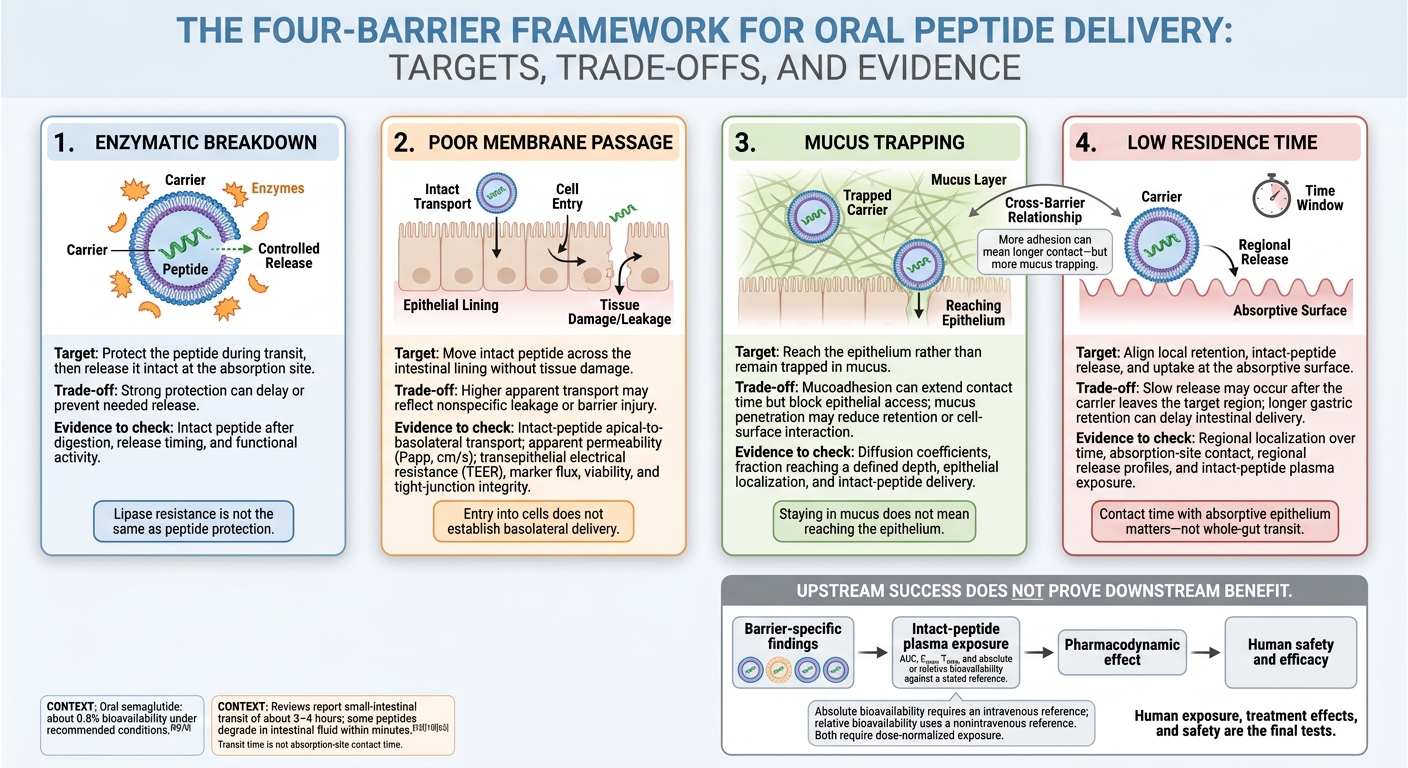
4 Barriers Lipid Carriers Target in Oral Peptides
Oral Peptide Delivery-Overcoming Bioavailability & Formulation Challenges | Thermo Fisher Scientific
sbb-itb-7164bd9
1. Enzymatic Breakdown
Pepsin, pancreatic proteases, and brush-border peptidases can destroy oral peptides. This vulnerability is a core challenge in peptide therapy. Encapsulation must protect the peptide during transit, then release it intact at the absorption site. Retention alone isn't enough - release timing matters just as much as protection.
Encapsulation, Lipid Digestion, and Release
Check where the peptide sits: in the lipid core, bilayer, or surface layer. Also assess lipid structure, surfactants, and leakage during storage or dilution. High initial encapsulation efficiency can hide early release, so measure release after dispersion and during the gastric-to-intestinal transition - not just at the final sampling point.
Bile salts and lipid digestion products can reorganize carriers into mixed micelles, vesicles, and other aggregates. These structures may keep the peptide dispersed, but they can also expose it to proteases too soon. Lipase resistance is not the same as peptide protection. A carrier that's harder to digest may also prevent the peptide from being released when needed.
The carrier's name matters less than how the formulation behaves during digestion.
| Carrier | Enzyme-relevant protection | Release behavior | Main limitation | Enzyme-stability findings |
|---|---|---|---|---|
| Liposomes | Limit direct enzyme access to encapsulated peptide | Leakage may occur before digestion; membrane composition can delay or trigger release | Physical instability and variable hydrophilic loading | Protection varies strongly with bilayer composition and surface properties. |
| SLNs | Reduce peptide diffusion and enzyme access | Often support slower or sustained release | Limited loading and lipid transitions can expel peptide during storage | Some tested SLNs aggregated in simulated gastric fluid at pH 2. Several showed no significant aggregation or disintegration in simulated intestinal fluid at pH 6.8, with or without trypsin. These findings measure carrier stability, not peptide integrity. |
| NLCs | Peptide incorporation may limit protease exposure | Can provide controlled release | Lipase digestion and surfactant exposure may destabilize the carrier | One insulin–sodium deoxycholate formulation retained approximately 30%–50% intact peptide after 4 hours of pancreatin exposure; the corresponding complex was completely degraded. |
| Lipid nanoparticles | Shielding depends on lipid chemistry and peptide location | May release peptide during digestion or retain it longer | Inconsistent terminology and incomplete identification of released species | Some systems improve stability, but direct comparisons remain limited. |
| Nanoemulsions | Lipid association may reduce direct protease exposure | Dilution, digestion, and reorganization can release peptide | Leakage can make release control difficult | Results depend on oil, surfactant, droplet size, and peptide location. |
Formulation-specific in vitro stability does not establish human exposure. Check intact-peptide plasma data separately.
Measuring Peptide Protection
Report pH, salts, bile components, enzyme identity and activity, temperature, mixing, fluid ratio, incubation time, and sampling intervals. Compare encapsulated peptide with free peptide, an empty carrier, and a physical mixture containing the same lipid dose. Run these comparisons both with and without enzymes.
Separate free peptide from carrier-associated peptide, quantify intact recovery, and identify fragments using selective chromatography with mass-spectrometric confirmation where appropriate. Missing controls, digestion conditions, or peptide-location data weaken comparisons.
Test the recovered peptide in a relevant functional assay, such as receptor binding or cell signaling, with free peptide as the reference standard. An intact chromatographic peak or a positive immunoassay doesn't necessarily mean the peptide still works.
In vitro protection, intact recovery, and functional activity do not establish systemic exposure. Check intact-peptide plasma concentrations, Cmax, Tmax, AUC, and absolute or relative bioavailability against a stated reference. Pharmacodynamic effects should match the peptide's mechanism. Neither simulated-fluid stability nor an animal response proves human systemic delivery.
Assess membrane passage only after establishing enzyme protection.
2. Poor Membrane Passage
After digestion is controlled, the next barrier is passage across the epithelium. Large, hydrophilic peptides do not readily partition into enterocyte membranes, and tight junctions limit paracellular passage. Protection from enzymes does not solve the permeability problem: an intact peptide still needs to cross the epithelial layer. This challenge comes before choosing a transport route, testing uptake, or assessing safety. For practitioners, practical guides on handling and administration provide additional context for clinical translation.
Epithelial Uptake and Transport
Carriers must reach the epithelium before uptake can occur. Endocytosis means entry into a cell; transcytosis also requires intracellular trafficking and basolateral exit. Judge each route by whether it moves intact peptide across the epithelium.
| Transport route | Relevant formulation features | Models | Safety or interpretation concern |
|---|---|---|---|
| Transcellular diffusion | Increased peptide lipophilicity or membrane-fluidizing agents | Artificial membranes, epithelial monolayers, intestinal tissue | Poorly suited to most intact hydrophilic peptides; membrane damage can make transport appear higher |
| Endocytosis | Particle size, surface charge, lipid composition, targeting ligands | Intestinal cells, microscopy, uptake assays | Entry into cells does not establish basolateral delivery |
| Receptor-mediated transcytosis | Ligands directed at enterocyte receptors or M cells | Receptor-expressing cells, organoids, ex vivo intestine, regional intestinal models | Receptor expression varies by region and species; binding alone does not prove intact-peptide delivery |
| Paracellular transport | Enhancers that alter tight junctions | Monolayers with electrical-resistance testing, marker flux, junction imaging | Increased passage may reflect nonspecific leakage or tissue injury |
To separate particle effects from excipient effects, compare free peptide, empty carrier, peptide-loaded carrier, and a physical mixture of peptide plus excipient. When testing targeting, include carrier controls with and without the targeting ligand. Higher transport accompanied by marker leakage suggests nonspecific barrier opening.
Lymphatic transport is a separate claim. Support it with lymph sampling, tissue distribution, or pharmacokinetic data.
A proposed mechanism must translate into intact-peptide transport without barrier injury.
Measuring Transport and Epithelial Safety
Report apparent permeability (Papp, in cm/s) for intact peptide - not just a fluorescent signal. State the exposed area, donor concentration, sampling time, and transported amount. Normalize uptake to cell number or protein, dose, and time, and remove or separately measure surface-bound material.
Confirm apical-to-basolateral delivery in mucus-containing models or intestinal tissue. If claiming site-specific release, compare duodenal, jejunal, ileal, or colonic uptake. Smaller particles and higher cellular uptake do not establish better absorption in humans.
Report transepithelial electrical resistance (TEER) before exposure, during treatment, and after washout. Pair these measurements with paracellular marker flux, viability, and tight-junction integrity. Persistent resistance loss, marker leakage, or tissue injury weakens the safety claim; recovery alone is not a complete safety assessment. Report intact-peptide plasma exposure separately from in vitro transport data.
3. Mucus Trapping
After digestion is controlled, mucus becomes the next barrier to epithelial access. Intestinal mucus can trap lipid carriers before they reach the epithelium. For oral peptides, it acts as a gatekeeper between stability and delivery. Staying in mucus does not mean reaching the epithelium.
Mucoadhesion vs. Mucus Penetration
Mucoadhesive surfaces extend contact time; mucus-penetrating surfaces improve diffusion.
| Design | Surface modification | Mucus behavior | Possible absorption advantage | Failure mode |
|---|---|---|---|---|
| Mucoadhesive | Cationic, hydrophobic, thiolated, or adhesive polymer surface | Binds mucins | Longer local contact for peptide release | Trapping in outer mucus or loss with mucus turnover |
| Mucus-penetrating | Dense hydrophilic PEG or zwitterionic coating | Reduces nonspecific binding | Better epithelial access for the peptide payload | Rapid transit, insufficient uptake, altered release, or reduced cell-surface interaction |
| Staged | Controlled adhesive and penetrating surface regions | Penetrates before retaining or releasing payload | Access followed by local contact | Complex manufacturing and uncertain in vivo behavior |
PEGylated solid lipid nanoparticles moved through mucus-producing models more effectively than unmodified particles, but the effect was formulation-specific and does not prove universal benefit.
Better carrier movement matters only if it also improves access for the peptide payload.
Measuring Mucus Movement and Epithelial Access
Report diffusion coefficients, the fraction reaching a defined depth, and retention or detachment during washing or fluid exchange. Specify the mucus source, temperature, medium, species, fed or fasted state, and gastrointestinal region. Measure epithelial localization separately from the bulk mucus signal, and confirm delivery of intact peptide. When reporting mucus turnover, include the source, model, region, and method. Do not treat one human small-intestinal turnover time as a general rule.
Distinguish carrier-associated signal, released peptide, and intact peptide. Pair imaging with validated measurements of peptide integrity, then link epithelial access to intact-peptide plasma exposure. Movement through mucus alone is not enough: some lipid carriers cross mucus and enter cells without improving transport of their payload.
Even when a carrier penetrates mucus, it may not stay at the absorption site long enough.
4. Low Residence Time
After protection, membrane-transport support, and mucus passage, a carrier still needs enough time at the absorptive surface to release intact peptide. Contact time with absorptive epithelium matters - not whole-gut transit. Reviews report small-intestinal transit of about 3–4 hours, but some peptides degrade in intestinal fluid within minutes.
Retention and Release at the Absorption Site
The goal is to align retention, release, and uptake, rather than simply slow transit. Food can delay gastric emptying and alter lipid digestion, which may delay intestinal delivery or change release timing. Test release under both fasted and standardized fed conditions. More time in the stomach may delay intestinal delivery rather than improve it.
| Strategy | Intended benefit | Possible failure mode | Data needed to show improved absorption |
|---|---|---|---|
| Mucoadhesion | Extend local contact for intact-peptide delivery | Adhesion can prevent the carrier from reaching absorptive epithelium | Localization over time, intact-peptide availability at the epithelium, and plasma exposure |
| Controlled release | Maintain intact-peptide availability during contact with the absorption site | Release may happen after the carrier leaves the target region; slow or incomplete release can reduce the available dose | Regional release profiles, intact-peptide mass balance, and plasma exposure |
| Lipid-digestion-triggered release | Align intact-peptide release with intestinal delivery | Food-dependent timing, premature burst release, peptide degradation, or carrier destabilization | Fed and fasted digestion studies, intact-peptide recovery, and bioavailability |
| Regional targeting | Release where epithelial uptake is most favorable | Changes in gastric emptying and motility may shift release outside the intended region | Regional imaging or sampling, transit measurements, and intact-peptide exposure |
Longer retention matters only when it increases intact-peptide delivery to absorptive tissue. Carrier fluorescence alone does not prove better peptide absorption. Include a same-dose comparator that is non-retentive or releases peptide quickly to help separate residence-time effects from other formulation effects. Tie residence-time claims to regional exposure and intact-peptide absorption.
Measuring Residence Time and Absorption
Where feasible, measure gastric emptying, intestinal transit, regional localization, and absorption-site contact separately. Imaging, gastrointestinal sampling, and tissue measurements show location directly; delayed plasma appearance offers only an indirect clue. Report where the peptide is - not just the lipid label. Use an assay that distinguishes intact peptide from fragments, with mass spectrometry to confirm identity.
Pair regional delivery measurements with intact-peptide AUC, Cmax, and Tmax, alongside absolute or relative bioavailability. Also assess pharmacodynamic onset and duration against therapeutic effect under fed and fasted conditions. For more on translating these findings into practice, see our clinical insights on peptide therapy. A later Tmax may reflect delayed emptying or slower release, and Cmax can fall without improving AUC. A longer effect does not prove longer residence. Match regional delivery, intact-peptide exposure, and therapeutic response.
How to Assess Published Lipid Carrier Studies
Use the four-barrier framework to judge what a study proves. Rate each study by the highest barrier it demonstrates - not by how promising the carrier sounds. Encapsulation, fluorescence, uptake, and apparent permeability do not prove systemic delivery or clinical benefit. Keep every claim within the evidence shown.
From Formulation Tests to Human Studies
Start with formulation quality and stability. Review assay methods, replicate measurements, acceptance criteria, and peptide recovery after simulated gastrointestinal exposure. Do the digestion conditions match the barrier being claimed? Studies should include both free-peptide and empty-carrier controls.
For epithelial studies, separate surface binding and intracellular uptake from intact peptide reaching the receiver compartment. Check mass balance, cell viability, and barrier integrity.
Absolute bioavailability requires an intravenous reference; relative bioavailability uses a nonintravenous reference. Both require dose-normalized exposure. For human studies, also review patient selection criteria, randomization, sample-size justification, adherence, meal conditions, confidence intervals, and tolerability. Beating free peptide can still mean low absolute exposure.
Even strong methods may support only a limited claim when the endpoint stops short of intact-peptide exposure.
Mixed Results and Safety Limits
Lipid carriers do not perform equally across barriers, and no single feature addresses all four limits. Improving one barrier can come at the expense of another: adhesion can increase mucus trapping, smaller size may not improve absorption, protection can delay release, and triggered release can happen too early.
Before crediting one feature for a result, compare composition, surface chemistry, and model conditions. Cell, tissue, animal, and human findings often differ, so keep each result within its own evidence tier.
Single-dose tolerability is not enough for chronic treatment. Look for repeated-dose evidence on GI injury, inflammation, immune effects, and excipient toxicity. Check manufacturing consistency, too. Excipients should have a regulatory history for the intended oral dose - not just for another route.
Conclusion: Match Each Barrier to the Data
Lipid carriers can help address enzymatic breakdown, membrane passage, mucus trapping, and residence time. Test each claim with a barrier-specific endpoint: intact peptide after digestion, epithelial transport, mucus penetration, or retention plus release.
For all four barriers, match the endpoint to the claim. Connect carrier properties to barrier behavior, intact-peptide exposure, pharmacodynamic effect, and clinical benefit. Upstream success does not prove downstream benefit.
Human data - not formulation success - determine clinical value. Clinical adoption requires reproducible human exposure, an exposure-response relationship, acceptable variability, and safety and efficacy at the intended dose. Select the carrier based on the barrier, and assess the therapy through human exposure, safety, and efficacy.
FAQs
How can I tell which barrier limits a peptide’s absorption?
Look for tests that measure each barrier separately: peptide stability in simulated gastric or intestinal fluids for enzymatic breakdown; TEER or FITC-dextran tracer assays for membrane passage and leakage between cells; and labeled-tracer retention measurements over time for mucus trapping and residence time.
Whole-tissue concentrations can be misleading. Check whether the data separate local accumulation from systemic clearance - and buildup between cells from actual uptake into cells.
Can one lipid carrier address all four barriers?
No single lipid carrier currently tackles all four barriers: enzymatic breakdown, poor membrane passage, mucus trapping, and low residence time. Lipid-based systems improve stability and transport, but dose standardization and complex biological barriers remain challenges.
Addressing these barriers often means pairing charge-based targeting with structural protection, such as nanoparticle encapsulation or hydrogel depots for sustained release. Limited human data still restrict clinical adoption.
What evidence would justify switching to an oral peptide?
Before switching, look for strong clinical evidence, ideally from multiple human randomized controlled trials or large-scale meta-analyses. Prioritize FDA-approved options, and verify that the specific oral formulation has shown bioavailability and efficacy in humans. Animal studies or theories about how it might work aren’t enough on their own.
Document each patient’s needs and set up a monitoring plan. Confirm that the product comes from a licensed 503A or 503B pharmacy that provides third-party Certificates of Analysis for purity and potency.