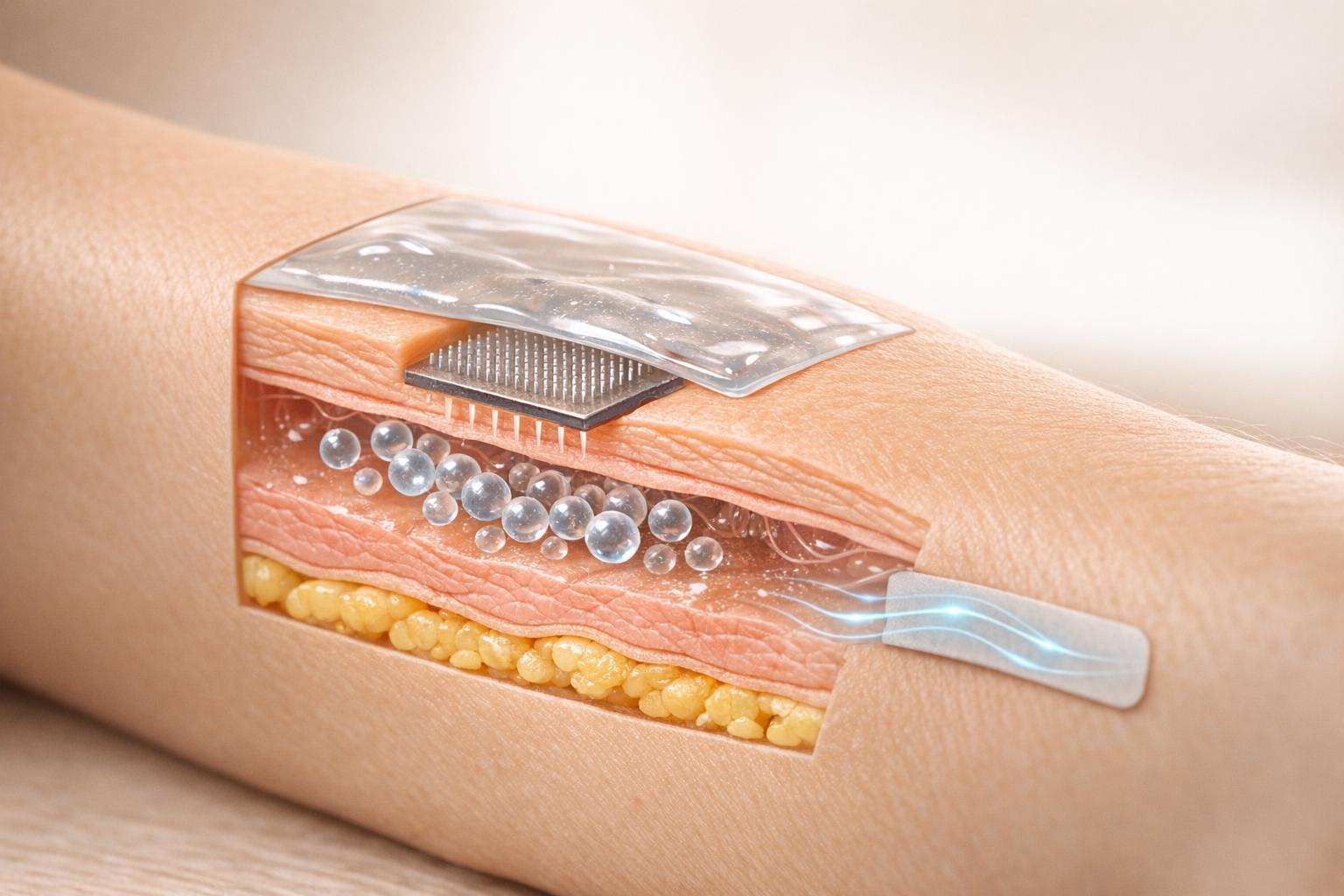
Peptide delivery through the skin faces major challenges due to the skin's natural barrier, the stratum corneum, which blocks most hydrophilic and large molecules. Traditional injections, while effective, are often uncomfortable for patients. This article explores non-injection methods like liposomes, hydrogels, microneedles, and iontophoresis to improve peptide delivery. Here's a quick breakdown:
- Liposomes: Phospholipid-based systems that enhance peptide penetration. Advanced types like transferosomes and ethosomes improve delivery into deeper layers.
- Hydrogels & Polymers: Offer controlled release and are effective for wound healing and soothing sensitive skin.
- Microneedles: Create tiny skin channels for direct delivery, reducing discomfort and improving bioavailability.
- Iontophoresis: Uses mild electric currents to push peptides through the skin, effective for smaller molecules.
Each method has strengths and limitations, making it important to match the system to the clinical goal. For example, microneedles are ideal for deep delivery, while hydrogels work well for surface treatments. The article also highlights clinical considerations, safety profiles, and practical considerations for these systems.
Deeper Clinical Results: Novel Delivery Systems for Protein & Peptide-Based Actives
sbb-itb-7164bd9
1. Liposomal and Vesicular Systems
Liposomes are tiny, phospholipid-based vesicles that naturally interact with skin lipids, making them effective for delivering peptides into deeper layers of the skin. Vesicular systems, however, differ in how well they penetrate, perform, and how convenient they are to use. Let’s dive into some key aspects, starting with how these systems penetrate the skin, alongside other clinical insights for practitioners.
Depth of Penetration
Traditional liposomes mostly stay within the upper layer of the skin, the stratum corneum. To address this limitation, advanced systems like transferosomes and ethosomes have been developed.
- Transferosomes: These use edge activators, such as surfactants like Tween 80 or Span 80, to make their membranes ultra-flexible. This flexibility allows them to compress and pass through tiny intercellular spaces, guided by the skin's natural hydration gradient.
- Ethosomes: These vesicles include 20–45% ethanol, which disrupts the tightly packed lipid matrix of the stratum corneum. Ethanol also increases the fluidity of the vesicle membranes, enhancing penetration.
The difference in performance between these advanced systems and conventional liposomes is striking. For instance, transferosomes carrying ketotifen fumarate delivered 3 times more drug into the epidermis and dermis compared to standard liposomes. Similarly, phosphatidylcholine–sodium cholate transferosomes achieved over 5 times greater penetration of diclofenac than conventional liposomes.
Clinical Effectiveness
The potential of liposomal systems is highlighted by their ability to deliver large peptides like Botulinum toxin-A (BTXA), which has a molecular weight of 70 kDa. A clinical study published in July 2024 in the Journal of Materials Science: Materials in Medicine showed that phosphatidylcholine/cholesterol nanoliposomes delivered BTXA to a depth of 380 μm - 8 times deeper than pure BTXA, which only reached 50 μm. In a trial involving 12 patients with post-acne facial scarring, combining liposomal BTXA with a hyaluronic acid hydrogel achieved a 91.67% total effective rate, compared to just 66.67% for the hyaluronic acid hydrogel alone.
In anti-hyperpigmentation treatments, anionic tripeptide-loaded liposomes (CSF sequence) demonstrated impressive results in a June 2025 study. These liposomes achieved an epidermal retention of 4.65 ± 1.81 μg/cm² in ex vivo human skin tests, effectively targeting melanocytes without any systemic absorption detected. These findings highlight the potential for safe and effective peptide delivery in non-invasive treatments.
Safety and Tolerability
Liposomal systems are generally well-received by the skin. Studies using reconstructed human epidermis (RHE) models found near 100% cell viability with anionic tripeptide liposomes, confirming their biocompatibility and the biodegradable nature of phospholipids.
"The prepared carriers were found to be stable and exhibited no cytotoxicity toward reconstructed human epidermis cells." - Michał Dymek, Faculty of Chemical Engineering and Technology, Cracow University of Technology
Anionic formulations are favored over cationic ones for long-term use, as cationic vesicles are more likely to cause skin irritation.
Practicality in Clinical Use
Despite their promise, challenges remain in bringing these systems into widespread clinical use. Ensuring manufacturing consistency and stability is a key hurdle. Advanced microfluidic techniques can produce ultrasmall nanoliposomes (below 40 nm) with a polydispersity index (PDI) under 0.2, ensuring consistent quality across batches. However, these methods require specialized equipment, which is not standard in many clinical settings.
To maintain stability during storage and transport, lyophilization (freeze-drying) is the most reliable method. Additionally, integrating vesicles into microneedle arrays (discussed in Section 3) could address some of the current limitations, offering a new way to enhance delivery methods in clinical applications.
2. Hydrogel and Polymer-Based Systems
Hydrogel and polymer-based platforms take a different approach from lipid-based systems, relying on hydrophilic matrices and engineered polymers to regulate peptide release. These systems address some of the limitations of liposomal systems, offering distinct advantages and trade-offs in areas like penetration depth, release timing, and ease of clinical use.
Depth of Penetration
Like other topical solutions, standard hydrogels face challenges in penetrating the stratum corneum. Typically, less than 1–5% of the applied dose reaches the viable layers of the epidermis and dermis. To overcome this, researchers have explored two approaches: enhancing hydrogels with cell-penetrating peptides (CPPs) or using polymer-based microneedle systems to bypass the skin barrier altogether.
CPPs such as TAT, when integrated into hydrogel formulations, significantly improve the permeability of large proteins through the skin. For instance, in wound-healing studies, hydrogels containing TAT-conjugated human growth hormone (hGH) achieved complete wound recovery by day 8, while control groups still showed 10% of the wound unhealed. On the other hand, polymer-based microneedle patches, particularly those using PLGA, address the barrier issue mechanically, delivering peptides directly into the dermis without relying on chemical modifications.
Clinical Effectiveness
The release characteristics of these systems set them apart. Peptide-enriched hydrogels release 75–80% of their payload within hours, making them ideal for applications like surface repair, hydration, and calming irritated skin.
In contrast, PLGA-based microneedle patches offer extended release. A study led by Kun Cheng at the University of Missouri-Kansas City, published in ACS Biomaterials Science & Engineering in December 2025, demonstrated an emulsion-based PLGA microneedle system capable of delivering peptides over 72 hours in vivo while maintaining their biological activity during storage. This sustained release is a clear advantage over traditional hydrogels.
Similarly, in April 2026, Scientific Reports highlighted a peptide-enriched hydrogel developed by the Medical University of Gdańsk (NE1 and IM2). This hydrogel was shown to be non-irritating, effective at relieving itching and tension, and compatible with microbial purity and packaging standards. These contrasting release profiles emphasize the importance of aligning the delivery system with the specific clinical need and patient selection criteria.
Safety and Tolerability
When properly formulated, both hydrogel and polymer-based systems demonstrate strong safety records. Natural polymers like hyaluronic acid and chitosan are not only safe but also promote skin regeneration, showing minimal cytotoxicity and low immunogenicity. PLGA, being FDA-approved, adds a layer of regulatory assurance.
"The cosmetic composition is stable, possesses preservative properties, and is safe in both in vitro and in vivo studies." - Scientific Reports
Hydrogels also have a practical edge in patient adherence. Their cooling and soothing properties make them more comfortable for sensitive or compromised skin, which is particularly valuable in conditions like atopic dermatitis, a condition affecting about 20% of children worldwide.
Practicality in Clinical Use
Here’s a snapshot of the practical differences between the main systems:
| System Type | Release Profile | Key Clinical Advantage | Main Challenge |
|---|---|---|---|
| Peptide-Enriched Hydrogel | Rapid (75–80% in first hours) | Soothing, suitable for sensitive skin | Requires preservatives for stability |
| PLGA Microneedle Patch | Sustained (up to 72 hours) | Deep dermal delivery; FDA-approved material | Requires organic solvents for fabrication |
| Lipid-Polymer Hybrid (LPHN) | Tunable | Balances stability and biocompatibility | Complex manufacturing and scaling issues |
While these systems hold great clinical promise, scaling them up for widespread use remains a hurdle.
"Gaps persist: scaled-up production, physical instability of the formulation, regulatory testing, and low level of clinical adoption." - International Journal of Peptide Research and Therapeutics
Microfluidic mixing is emerging as a promising manufacturing method for hybrid polymer systems, offering improved batch consistency. However, the specialized infrastructure required for this process is not yet widely available in clinical settings.
Next, we’ll explore microneedling techniques that further refine peptide delivery to the dermis.
3. Microneedling and Microneedle Patches
Microneedling offers a game-changing approach by bypassing the limitations of surface-based delivery systems. While hydrogels and polymer systems work on the skin's surface, microneedling goes deeper, creating direct pathways through the skin without relying on chemical enhancers. Unlike liposomal or hydrogel-based methods that depend on chemical aids, microneedle patches physically breach the skin barrier, providing a more direct route for delivery.
Depth of Penetration
Microneedle systems are designed to penetrate the skin at varying depths, depending on their type. Standard solid microneedling devices create microchannels about 50–100 µm deep, which is sufficient for peptides to diffuse through. Dissolving and coated microneedle patches go further, reaching depths of 100–600 µm, allowing them to deliver their payload directly into the epidermis or upper dermis. For even deeper delivery, hollow microneedle systems can penetrate up to 2,000 µm, making them ideal for higher-volume biologic drugs.
"MN arrays consist of microscopic projections (typically 100–1000 μm in length) that penetrate the stratum corneum to create transient microchannels in the epidermis or upper dermis without stimulating pain receptors or blood vessels." - AAPS PharmSciTech
These microneedle arrays efficiently bypass the stratum corneum by creating temporary channels. However, due to the skin's viscoelastic nature, needles often don’t reach their full rated depth. Even so, the effective penetration achieved is enough to support strong clinical outcomes.
Clinical Effectiveness
Microneedle patches are gaining traction in clinical applications. A study published in Science Translational Medicine in November 2024 by Zhen Gu and Yuqi Zhang at Zhejiang University demonstrated the potential of wearable osmotic microneedle (OMN) patches. These patches delivered the peptide drug exenatide continuously for 24 hours in murine models, maintaining stable plasma levels. By comparison, subcutaneous injections resulted in a rapid spike followed by a steep decline. In human trials, participants found the OMN patch comfortable and preferred it over traditional methods.
The bioavailability of microneedle systems is another key advantage. Research published in AAPS PharmSciTech in 2026 showed that dissolving microneedle patches loaded with liraglutide, a GLP-1 agonist, achieved 69.8% relative bioavailability in rats and 46.3% in minipigs compared to subcutaneous injections. These results are impressive, especially when you consider that oral peptide delivery often loses most of its effectiveness due to degradation in the gastrointestinal tract and first-pass metabolism.
Safety and Tolerability
Microneedle systems are generally safe for most patients. The microchannels they create are temporary, starting to close within 30 minutes and fully healing within 6 hours. The skin barrier typically recovers completely within 1–2 days, which greatly reduces the risk of infection.
Dissolving patches, made from biodegradable materials such as hyaluronic acid, PLGA, or carboxymethyl cellulose, leave no sharps waste and carry minimal risk of systemic side effects. These patches are also a great option for the 18–40% of people who have a fear of needles.
Practicality in Clinical Use
Microneedle systems offer practical advantages beyond their clinical effectiveness and safety. One standout feature is their ability to enhance peptide stability. For instance, parathyroid hormone (PTH) coated on microneedles remained stable for two years at 77°F (25°C), potentially removing the need for cold-chain storage. This could be a game-changer for clinics managing multiple peptide therapies, simplifying logistics and reducing costs. For more on implementation, see our practical clinical guides.
| Microneedle Type | Mechanism | Depth | Benefit |
|---|---|---|---|
| Solid | Poke-and-patch | 50–100 µm | Cost-effective; reusable |
| Dissolving | Poke-and-release | 100–600 µm | No sharps waste; stable storage |
| Hollow | Poke-and-flow | Up to 2,000 µm | Precise delivery for high-volume biologics |
| Osmotic | Osmotic pressure | Variable | Continuous 24-hour delivery; no electronics required |
Despite these benefits, regulatory hurdles remain a challenge. Microneedle products are classified as drug-device combination products, and quality control standards are still evolving to keep pace with the technology.
4. Iontophoresis and Energy-Based Methods
While microneedle patches physically penetrate the skin, iontophoresis offers a different route. It uses a mild electrical current to push peptides through natural low-resistance pathways like hair follicles and sweat ducts. Unlike passive topical treatments that rely on diffusion, iontophoresis actively drives delivery.
Depth of Penetration
Iontophoresis works through two key mechanisms: electromigration (repelling charged molecules) and electroosmosis (moving bulk fluid). This approach allows the delivery of larger peptides, such as teriparatide (~4,000 Da) and insulin (~5,800 Da), which passive methods can't handle. However, it struggles with peptides above 13 kDa on its own. Combining iontophoresis with microneedling addresses this limitation, as shown by a 17-fold increase in ovalbumin delivery compared to passive diffusion. This synergy highlights its potential for better clinical outcomes. Clinicians often integrate these methods into standardized peptide dosing protocols to ensure safety and efficacy.
Clinical Effectiveness
Iontophoresis expands the range of peptides that can be delivered, including those up to 5,800 Da, offering more possibilities for treatment. In January 2026, Ryuse Sakurai and his team at the Tokyo University of Science tested pulsed-current iontophoresis for delivering teriparatide in rats. After 2 hours at 0.3 mA/cm², serum concentrations reached 53.3 ± 4.0 pg/mL - on par with constant direct current but with reduced skin damage. For smaller peptides, such as the anti-inflammatory peptide KPV, iontophoresis at 0.3 mA/cm² increased skin retention by 10 times compared to passive methods, where diffusion was nearly undetectable.
"Pulsed-current IP enables noninvasive and effective systemic delivery of peptide drugs with minimized skin irritation, representing a promising alternative to injection-based administration for macromolecular therapeutics." - Ryuse Sakurai, Faculty of Pharmaceutical Sciences, Tokyo University of Science
Safety and Tolerability
While iontophoresis is effective, managing skin irritation is critical. The type of current plays a big role here. Pulsed current at 10 Hz keeps transepidermal water loss (TEWL) at about 15.8 g/m²/h, reducing erythema and edema. In contrast, constant direct current significantly disrupts the skin barrier, with TEWL jumping from around 5 to 47.5 ± 8.7 g/m²/h after 50 hours. To minimize irritation, it's recommended to keep current density below 0.5 mA/cm² and favor pulsed modes over constant direct current. Clinicians should also monitor delayed skin reactions, as edema can peak up to 26 hours post-treatment.
Practicality in Clinical Use
Recent innovations have made iontophoresis more practical. In May 2024, xTrans Creative Inc. and the National Atomic Research Institute in Taiwan unveiled a self-powered patch using printed Zn/Ag electrodes. These electrodes generate a microcurrent (0.6–0.74 V) upon contact with biological electrolytes, increasing the penetration of acetyl hexapeptide-8 by 1.5 times compared to standard patches. The patch also drove fluorescein to a depth of 0.4 mm within 20 minutes. Additionally, lightweight wearable systems - some weighing just 18 grams - make self-administration at home a realistic option.
| Feature | Constant DC Iontophoresis | Pulsed-Current (10 Hz) | IP + Microneedles |
|---|---|---|---|
| Peptide Size Limit | Typically <10–13 kDa | Typically <10–13 kDa | Large macromolecules (>13 kDa) |
| Skin Irritation | High; TEWL spikes | Significantly reduced | Low; minimally invasive |
| Delivery Efficiency | Effective | Comparable to DC | Superior for large peptides/vaccines |
| Mechanism | Electrorepulsion/Osmosis | Intermittent depolarization | Physical puncture + active drive |
Pros and Cons
Every delivery system has its own strengths and limitations, making it important to weigh these factors carefully in clinical settings. Below is a comparison of key features across different delivery methods, focusing on penetration depth, clinical outcomes, safety, and ease of use.
| Delivery System | Penetration Depth | Clinical Outcomes | Safety | Ease of Use |
|---|---|---|---|---|
| Liposomes | Limited (mainly epidermis retention) | High; backed by clinical validation | Biocompatible; non-cytotoxic | High; suitable for topical use or peptide injection |
| Transferosomes | Deep; can reach systemic circulation | Moderate; primarily preclinical | Generally safe; minor irritation possible from surfactants | High; easy topical application |
| Hydrogel/Polymer Systems | Moderate to high | Sustained release up to 72 hours; effective for wound healing | High; uses biocompatible materials | High; often comes in patch form |
| Dissolving Microneedles | Deep (epidermis/dermis) | High; comparable to subcutaneous injection | High; eliminates sharps waste and lowers infection risks | High; can support self-administration |
| Hollow Microneedles | Deep (dermis) | High; allows precise dose control | Moderate; risks include pain and clogging | Low; requires additional devices or pumps |
| Iontophoresis | Moderate; up to ~13 kDa alone | Effective for charged peptides; better with microneedles | Moderate | Low; relies on specialized equipment |
This comparison sheds light on the clinical applications and limitations of each system. For example, liposomes are well-validated for localized delivery but struggle to achieve systemic effects due to limited skin penetration. On the other hand, dissolving microneedles stand out for their versatility, offering easy self-administration, reduced needle-stick risks, and stable peptide storage at room temperature (up to two years at 77°F/25°C).
Iontophoresis, while effective for certain charged molecules, requires specialized equipment and precise current control, making it more suitable as an adjunct to other methods. These distinctions are crucial for tailoring treatment strategies to individual patient needs.
Conclusion
Matching the right delivery system to a specific clinical goal can make a noticeable difference in treatment outcomes. The examples above highlight how tailoring delivery methods to meet both clinical objectives and patient needs improves the effectiveness of therapies.
For anti-aging treatments, transferosomes and HA microneedle patches stand out. Transferosomes deliver up to three times more drug to the epidermis and dermis compared to conventional liposomes, while HA microneedle patches offer a minimally invasive alternative. When it comes to wound healing and radiation-damaged skin, peptide-enriched hydrogels, especially those containing GHK-Cu sequences, are a practical choice, releasing 75–80% of their peptide content within the first few hours. For scar reduction, siRNA microneedle patches outperform traditional silicone sheets in postoperative care.
Patient characteristics also play a critical role. Hydrogels are ideal for individuals with sensitive or compromised skin. Dissolving microneedle patches address the needs of the 18–40% of people with needle phobia, while PLGA-based microneedles are suited for chronic or metabolic conditions, sustaining peptide release for up to 72 hours.
"MN-assisted delivery represents a promising strategy for patient-centered, stable, and accessible biologic therapies." - AAPS PharmSciTech
FAQs
Which delivery system is best for my treatment goal?
When choosing a delivery system, it all comes down to your clinical goals. If you're aiming for systemic benefits - like improved skin hydration or a brighter complexion - oral polypeptides tend to deliver better results. On the other hand, if your focus is on specific local concerns, such as reducing wrinkles or addressing sensitive skin, topical treatments are a solid choice.
For even better results, advanced delivery methods can make a big difference. Techniques like microneedles, skin-penetrating peptides, and hydrogels help boost absorption, bypass the skin's natural barriers, and deliver moisture exactly where it's needed most.
How deep do microneedles actually deliver peptides in real skin?
Microneedles work by forming tiny channels through the stratum corneum - the skin's primary barrier - to deliver peptides. These projections, which usually measure between 25 and 2,000 micrometers in length, penetrate the epidermis or upper dermis. This precise depth enables access to the dermal microcirculation, enhancing peptide absorption while steering clear of deeper pain receptors and blood vessels.
What safety risks should I watch for with iontophoresis or microneedles?
When considering iontophoresis or microneedles for delivering peptides, it's important to keep a few things in mind. Iontophoresis involves the use of specialized equipment and requires careful handling to minimize risks. On the other hand, hollow microneedles, while effective, may cause more discomfort compared to traditional injections.
Both methods face limited clinical use, largely due to a lack of comprehensive safety and efficacy data. For clinicians looking to navigate these challenges, PeptidePrescriber offers evidence-based resources designed to promote safe and compliant practices with these delivery techniques.