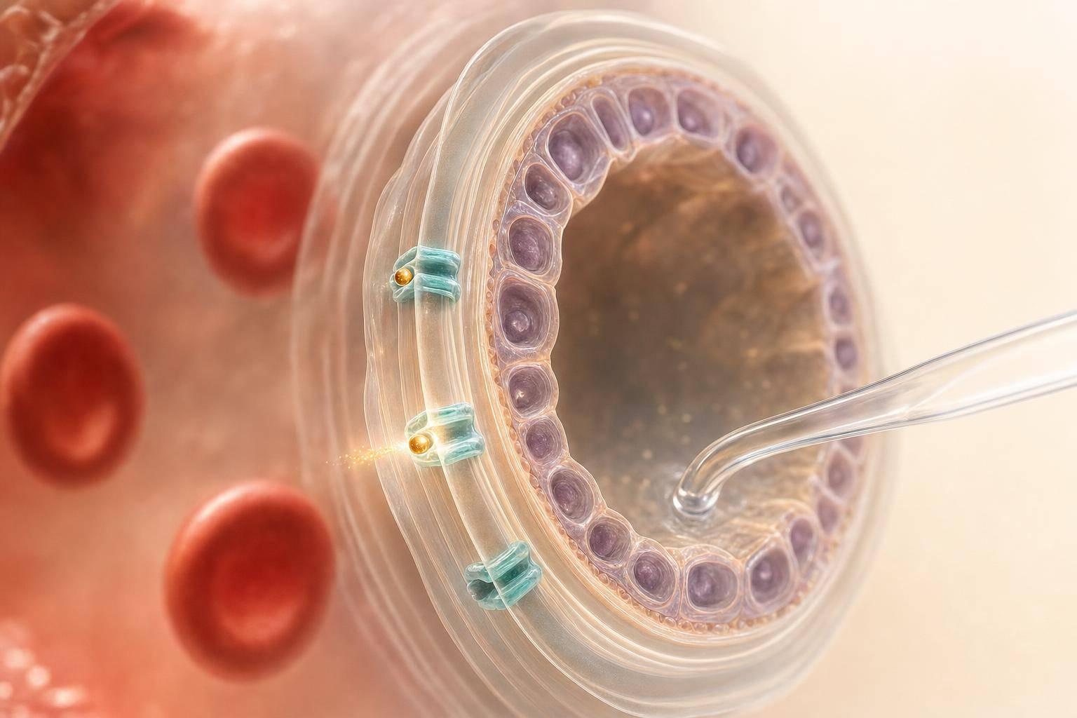
Most peptides do not freely cross the blood-testis barrier (BTB). If I’m reading a BTB paper, I focus on one question first: did the peptide reach the adluminal compartment behind an intact barrier, or did the study just show testis exposure, tissue retention, or barrier damage?
Here’s the short version:
- Whole-testis drug levels do not prove BTB crossing
- High TEER + low tracer leak support an intact barrier
- Influx and efflux transporters can change peptide entry and exit
- Barrier-opening studies show forced entry, not normal delivery
- Rodent data may not match humans because transporter location can differ by species
- Leydig cell effects can happen without BTB crossing because Leydig cells sit outside the barrier
- Luminal or rete testis sampling is more useful than bulk tissue levels when the goal is true intratubular exposure
A few data points matter right away. One cited rat study used a 22-amino acid occludin peptide at 0.4 or 4 μM and saw lower barrier function plus ¹²⁵I-BSA buildup in the treated testis. That shows barrier opening, not normal peptide transport. In older tracer work, inulin and [51Cr]EDTA were absent from rete testis fluid, which supports how tight the BTB is under normal conditions.
If I’m using this type of paper in practice, I read it in this order:
- Barrier status: Was the BTB intact?
- Transport pattern: Was movement directional, saturable, or blocked by inhibitors?
- Sampling site: Did the study separate interstitial tissue from adluminal fluid?
- Species fit: Do the transporters in the model match human biology?
- Clinical fit: Does the paper line up with reproductive tox, endocrine data, and human PK?
This gives me a simple way to separate true BTB passage from testis exposure that looks bigger than it is.
Blood testes Testis Barrier; formed by basal ectoplasmic specialization and gap junctions
sbb-itb-7164bd9
BTB Structure and Transport Mechanisms Relevant to Peptides
For peptide transport, the main question is simple: does the molecule reach the adluminal compartment, or does it stay on the basal side? The blood-testis barrier is formed by a junctional network between adjacent Sertoli cells, including tight junctions, adherens junctions, and gap junction parts such as connexin 43. That difference matters because changes in transporter activity or junction state can completely change what a paper reports as “testis uptake.”
Paracellular Restriction and Transcellular Movement
Under normal physiologic conditions, tight junctions between Sertoli cells place a strong limit on paracellular diffusion of peptides. So if a peptide is recovered from the adluminal compartment, that usually points to transcellular movement through Sertoli cells, not passive leak between them. In practice, that means uptake at the basal membrane, intracellular trafficking, and then release at the apical side into the adluminal space.
That’s why data on transcellular transport matter more than bulk tissue concentration alone. Uptake rates, intracellular accumulation, and apical release tell you far more about actual passage across the BTB than a single total testis concentration.
One thing to check in any BTB paper is whether the barrier was intact. Experimental occludin-derived peptides, for instance, can weaken Sertoli cell tight junctions. In rat studies, a 22-amino acid occludin peptide at 0.4 or 4 μM reduced tight-junction barrier function in bicameral culture, and intratesticular exposure produced ¹²⁵I-BSA accumulation in the treated testis compared with the untreated contralateral testis. That kind of result shows pathologic or drug-driven barrier opening, not normal physiologic delivery.
Influx and Efflux Transporters at the BTB
Junctions are only one part of the picture. Sertoli cell membranes also contain transporters that influence what gets into the testis and what gets sent back out. Basal uptake transporters - OCT, OAT, OCTN, and ENT1/ENT2 - may allow entry of peptide-like drugs, while efflux transporters such as ABCB1/P-gp, ABCC1/MRP1, and ABCA1 can limit adluminal accumulation.
When you read transport studies, it helps to look for two things:
- where the transporters were localized
- whether function was tested with inhibitor or substrate experiments
Without that, it’s hard to tell whether low testis exposure comes from junctional restriction, active efflux, or a mix of both. It also becomes harder to judge whether the measured signal reflects true passage across the barrier or just retention in tissue. These transporter effects also help explain why barrier permeability shifts during normal cycling and in disease.
Local Factors That Modify Transport
The BTB does not stay fixed. It remodels cyclically during spermatogenesis, which creates short-lived shifts in permeability. Endogenous regulatory peptides drive much of this remodeling. CNP can transiently loosen Sertoli-cell junctions and reopen the barrier after removal.
Inflammation adds another layer. Orchitis, systemic infection, or autoimmune processes can disrupt tight junctions, increase paracellular permeability, and change the expression or localization of both influx and efflux transporters. So a peptide that shows little testicular exposure in a healthy tissue model may reach higher concentrations when the barrier is compromised. That’s a big deal when reading disease-state studies, because the result may reflect an altered BTB rather than baseline transport behavior.
These structural features and local conditions are why different transport studies often rely on different models to measure BTB passage.
How BTB Peptide Transport Is Studied
Each model answers a different kind of question, so the first step is simple: figure out what you're trying to learn. If the goal is mechanism, researchers usually start in vitro. If the goal is exposure, they need in vivo or ex vivo sampling. And if the goal is to see whether the barrier can be opened on purpose, that calls for a different set of tools.
In Vitro Sertoli Cell and Transwell Models
For mechanism work, the standard setup uses primary or immortalized Sertoli cells grown on extracellular matrix-coated permeable inserts in a two-compartment culture system. After the cells form tight junctions, researchers place a peptide in one chamber and measure how much shows up in the other.
The main check for barrier integrity is transepithelial electrical resistance (TEER). If TEER rises and then holds steady, that's a sign the tight junctions have formed. Researchers usually back that up with tracer assays, often with fluorescent dextrans or radiolabeled inulin, to make sure paracellular diffusion is restricted before transport testing starts.
This setup is useful when the goal is to isolate what controls peptide movement. For example, it can help show which transporters or junction proteins matter, or how hormones, cytokines, or barrier-modifying compounds change transport.
That said, these models don't show how the intact organ behaves. In vitro systems are often leakier than the native BTB. They also leave out germ cells, peritubular myoid cells, and the 3D structure of a real seminiferous tubule. Human in vitro systems are usually leakier than rodent systems, so they're better for studying mechanism than for making direct clinical estimates.
In Vivo and Ex Vivo Testis Exposure Studies
Once the focus shifts from transporter behavior to actual tissue exposure, cell culture isn't enough. At that point, researchers move to whole-testis or luminal sampling. For clinicians, one distinction matters a lot: interstitial exposure is not the same as true BTB passage.
Whole-animal studies look at how much peptide reaches testicular tissue and how fast it gets there. Rete testis cannulation and seminiferous tubule microperfusion go a step further by collecting luminal fluid directly. That matters because it shows what crosses the BTB into the adluminal compartment, not just what builds up in interstitial tissue.
In classic permeability work, tracers like inulin and [51Cr]EDTA were absent from rete testis fluid, while smaller or more permeable substances entered more readily.
When reading these studies, a few metrics do most of the heavy lifting:
- Testis-to-plasma concentration ratio
- AUC in testicular tissue versus plasma
- Whether the sampling method separates interstitial exposure from true intratubular distribution
This is where interpretation can go sideways if you're not careful. A peptide that acts on Leydig cells may trigger a hormonal effect without ever crossing the BTB, because Leydig cells sit in the interstitium outside the barrier. Readouts tied to Sertoli cells or spermatogenesis are more useful when the goal is to judge actual BTB passage. In plain English: use these studies to tell apart interstitial exposure and true intratubular delivery.
Delivery Systems Designed to Increase BTB Passage
Some studies ask whether peptides pass through the BTB on their own. Others ask whether the barrier can be pushed open. Those are not the same thing.
Nanocarriers, including liposomes, polymeric nanoparticles, ferritin-based nanoreagents, and biomimetic frameworks, can use endocytic pathways in Sertoli or endothelial cells to move cargo across the barrier. Barrier-remodeling peptides can also transiently lower TEER and increase paracellular permeability.
The key point here is easy to miss: these results should be read as engineered penetration data, not as proof that routine systemic peptide dosing will cross an intact BTB. These are engineered penetration studies, not evidence that standard peptide dosing will cross an intact BTB.
How to Read BTB Transport Results in Research Papers
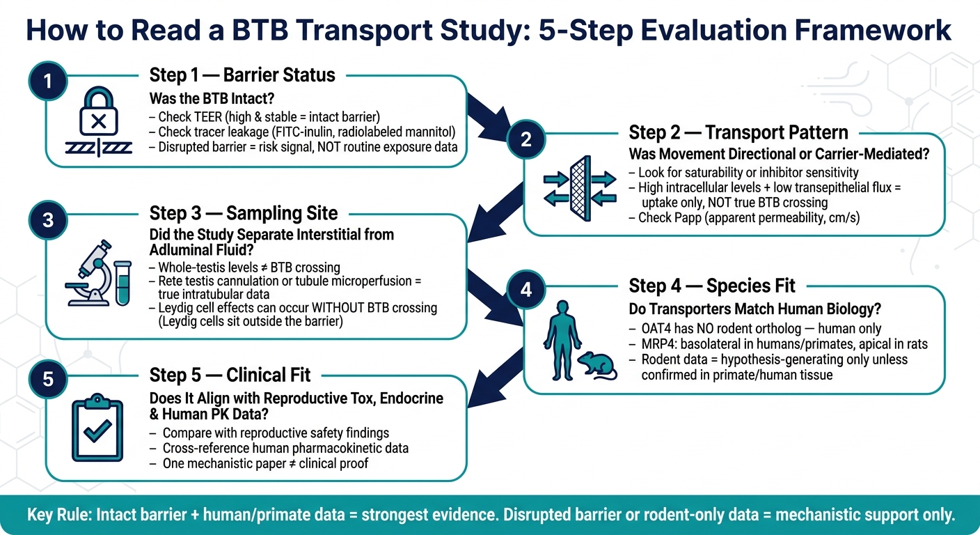
How to Read a Blood-Testis Barrier (BTB) Transport Study: 5-Step Framework
BTB transport papers are easy to misread if you jump straight to the headline result. A peptide can look like it moves well in a cell model, then look blocked in tissue, or end up in the wrong place because the transporter pattern changes by species.
A safer way to read these studies is to move step by step: barrier integrity first, then flux direction, then tissue distribution, and only after that species relevance.
Barrier Integrity and Permeability Terms
Before you trust any transport readout, check whether the barrier stayed intact.
TEER is the first thing to look at. It shows how tight the junctions are. If TEER stays high and stable during the experiment, that supports an intact barrier. But there’s a catch: Sertoli cell transwell models are leakier than the native BTB, so high permeability in culture does not automatically mean strong testis exposure in vivo.
Tracer leakage is the second check. If inert markers like FITC-inulin or radiolabeled mannitol show up in the receiver compartment, the barrier has broken down. At that point, any peptide signal is more likely due to paracellular leak than a real transport process.
Then comes apparent permeability (Papp), usually reported in cm/s. This tells you how fast a compound moves across the barrier. If you see higher directional Papp along with intact TEER, plus signs like saturability or inhibitor sensitivity, that points more toward carrier-mediated transport.
One detail gets missed all the time: High intracellular levels with low transepithelial flux suggest uptake without true BTB crossing. That matters a lot if the actual question is whether the peptide can reach germ cells, not just enter Sertoli cells.
Testis Uptake and Distribution Metrics
Once barrier integrity checks out, shift to exposure data. The key question here is simple: does the signal show true BTB passage, or is it just hanging around on the blood side?
The tissue-to-plasma ratio compares testis concentration with blood concentration at a given time point. A high ratio can look impressive, but it does not prove BTB crossing. Interstitial protein binding, vascular retention, or peritubular buildup can all push that number up even if the peptide never reaches the adluminal compartment.
The partition coefficient (Kp,testis) puts that same relationship into a steady-state frame. It can hint at slow tissue equilibration. Still, if the paper doesn’t separate interstitial from adluminal distribution, Kp,testis alone can’t tell you whether the compound was delivered to the adluminal side.
Concentration–time profiles like Cmax, Tmax, and AUC are most helpful when you read plasma and testis data together. Numbers from one tissue in isolation can send you down the wrong path. Studies are more convincing when they tie those pharmacokinetic results to functional readouts such as spermatogenesis, hormone levels, or local signaling, instead of stopping at tissue concentration alone. For more on translating these findings into practice, see our clinical insights on evidence-based therapy.
Species Differences and Translation Limits
After exposure, the next filter is species. This is where many BTB papers look stronger than they are.
Rodent and human BTB transporter localization can differ enough to change the whole readout. OAT4 (organic anion transporter 4) is a good example: it has no rodent ortholog and is found only in humans. So if a compound depends on OAT4, its behavior in a rat may tell you very little about what happens in a human.
MRP4 shows the same problem from another angle. In humans and non-human primates, MRP4 sits on the basolateral Sertoli cell membrane. In rats, it sits on the apical membrane. That one shift in location can change how you think about adluminal buildup for some anionic compounds.
Because of that, rodent BTB data should be treated as hypothesis-generating unless primate or human work shows the same transporter pattern. Clinicians looking to apply these findings can refer to our practical guides for administration and handling protocols.
Applying BTB Evidence in Clinical Practice
BTB transport findings are mechanistic, not clinical, conclusions. A rat study showing passage across the BTB can tell you how something may move. It does not tell you, by itself, that the same thing happens in patients, or that it matters clinically.
That’s the core idea here: read BTB findings alongside pharmacokinetics, reproductive toxicology, endocrine data, and human evidence. On their own, BTB studies only show part of the picture.
Key Checks When Reading a BTB Study
Before using a BTB paper to support a clinical decision, pause and run through six quick checks:
| Check | What to Look For | Why It Matters |
|---|---|---|
| Model type | In vitro cell system vs. in vivo animal | Not directly predictive |
| Species | Rodent vs. primate vs. human tissue | Species changes transporter localization |
| Route of administration | Intratesticular, intraperitoneal, subcutaneous | Limits translation |
| Peptide properties | Molecular weight, charge, lipophilicity | Determines plausibility |
| Transporter relevance | Which transporters were documented and under what conditions | Depends on species and barrier state |
| Barrier integrity | Was the BTB intact or deliberately disrupted? | Intact barrier required for transport claims |
That last check matters a lot. Some studies use chemical agents, inflammatory stimuli, or genetic manipulation to open up the BTB on purpose. Those studies can help us understand disease pathways and barrier failure. But they do not reflect the intact barrier setting of a healthy adult male patient.
A simple way to think about it: if the barrier had to be broken first, the finding is a risk signal, not a prediction of routine clinical exposure.
Once you’ve gone through those checks, compare the BTB data with reproductive safety findings and human exposure data. Don’t treat the BTB result as a standalone answer.
Using Structured Resources to Place BTB Data in Context
Structured clinical summaries can help when a single BTB paper starts to look bigger than it is. Use PeptidePrescriber monographs, safety summaries, and regulatory references to place BTB findings next to pharmacology, reproductive safety, and sourcing guidance.
That side-by-side view helps keep one mechanistic paper from driving the whole decision.
Conclusion: What BTB Transport Studies Can and Cannot Tell Clinicians
The BTB is one of the tightest blood–tissue barriers in the mammalian body. Whether a peptide crosses it depends on transporter biology, the peptide's own physicochemical properties, the state of the barrier, and the local microenvironment of the testis. No single study captures all of that completely.
So the practical read is pretty simple: give the most weight to intact-barrier human or primate data. Treat rodent studies and disrupted-barrier models as mechanistic support, not clinical proof.
FAQs
How can I tell if a peptide truly crossed the BTB?
Look for direct evidence that the peptide reached the target tissue, not just changes in bloodwork, hormone levels, or other body-wide markers.
Check the study methods for distribution data that shows where the substance actually went. A lot of peptide research comes from preclinical animal work or in vitro testing, so it helps to separate whole-body effects from actual tissue-level penetration.
Why don’t whole-testis drug levels prove BTB passage?
Whole-testis drug levels do not prove blood-testis barrier (BTB) passage. They only show the total amount of drug in the entire organ, not its exact location.
That distinction matters. The testis contains interstitial tissue, blood vessels, and seminiferous tubules. So a whole-testis measurement can't tell whether the drug is still sitting in the vasculature or whether it actually crossed the BTB and entered the protected tubular compartment.
How well can animal BTB data predict human exposure?
Animal blood-testis barrier (BTB) transport findings are only weak predictors of human exposure. In preclinical models, the evidence is usually less reliable, and animal results often don’t carry over cleanly to humans, especially when the delivery method, formulation, or dose is different.
If a transport study is preclinical only, treat it as hypothesis-generating rather than clinically predictive. When estimating exposure, lean on human PK/PD or controlled human data whenever possible.