
Early memory loss does not prove a leaky blood-brain barrier - or harmful brain inflammation. My main takeaway: if your memory is worsening, start with a clinical evaluation, not tests or supplements marketed for “BBB repair.”
Here’s how I’d frame the two pathways:
- Blood-brain barrier dysfunction affects what moves between your blood and brain.
- Microglial activation changes how the brain’s immune cells respond. That response can protect brain tissue or, if it persists, damage memory networks.
- Both can feed each other. Neither always comes first, and current tests cannot establish which started your symptoms.
I’d focus first on cognitive testing, daily function, medications, sleep, mood, and vascular health. A normal routine MRI does not rule out subtle barrier leakage, but specialized tests cannot prove it caused memory loss.
<u>No peptide or dietary supplement is established to repair the human BBB or treat mild cognitive impairment.</u> Follow-up is usually recommended within 6–12 months, or sooner if symptoms change. Alzheimer’s treatment may be an option for some patients after specialist assessment and confirmation of amyloid pathology.
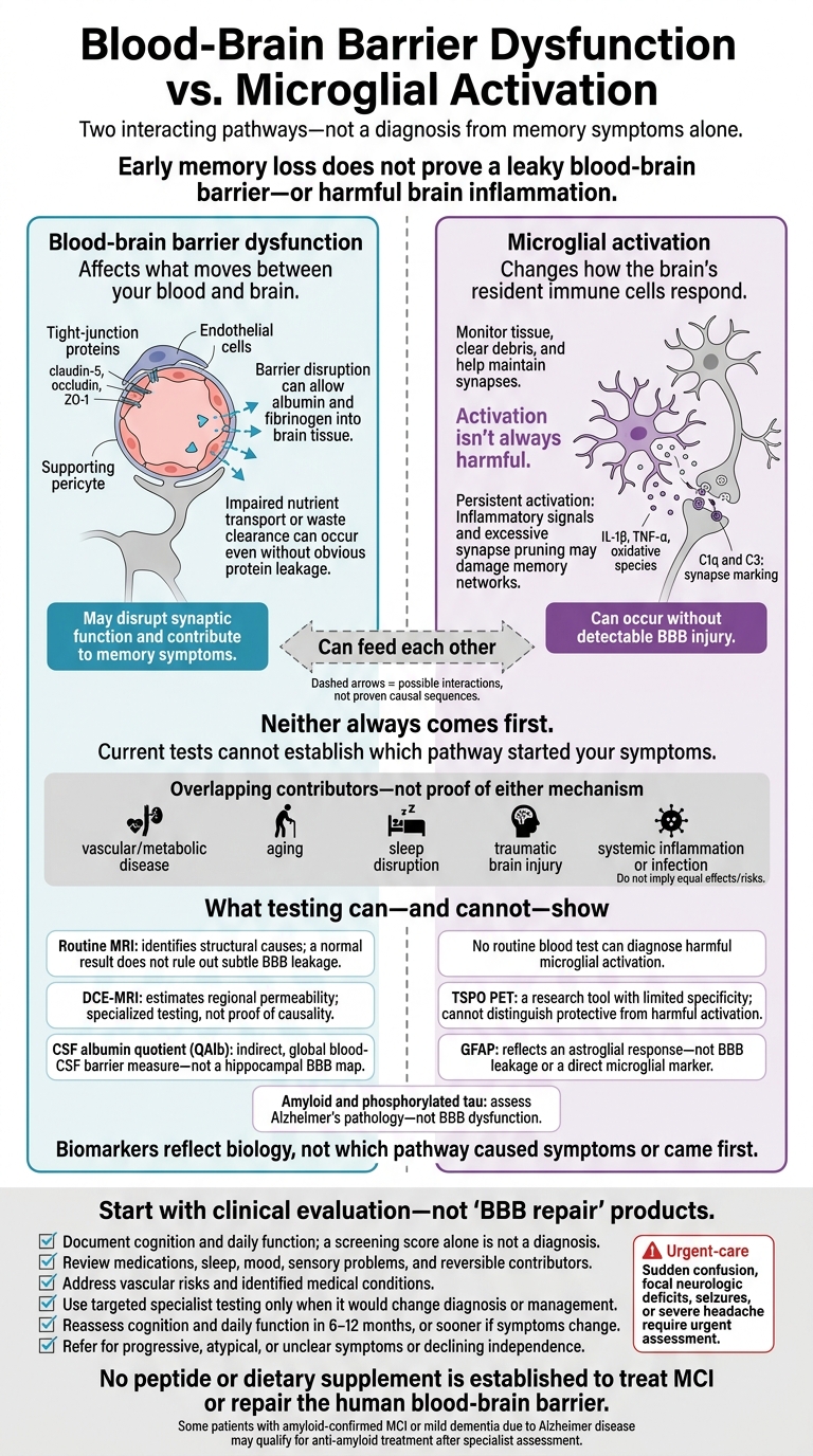
Blood-Brain Barrier Dysfunction vs. Microglial Activation
1. Blood-Brain Barrier Dysfunction
Mechanisms and Causal Evidence
Endothelial tight junctions (claudin-5, occludin, ZO-1) and pericytes help keep the blood-brain barrier (BBB) intact. When these fail, albumin and fibrinogen can enter brain tissue, fueling inflammation and disrupting synaptic function. BBB dysfunction can also involve impaired transport: nutrients may not reach brain tissue properly, or waste may not clear, even without obvious protein leakage.
BBB dysfunction may contribute to memory symptoms before changes appear on routine structural imaging. The hippocampus is especially vulnerable. Some studies found BBB breakdown before atrophy; others found permeability changes with or without abnormal amyloid or tau.
But findings differ across groups. A DELCODE/DESCRIBE analysis found hippocampal barrier disruption in Alzheimer’s dementia, but no statistically significant difference between participants with MCI and those with normal cognition. BBB injury may come before, occur alongside, or follow other pathology.
Testing Limits
In MCI, clinicians usually infer BBB injury rather than see it directly. A normal routine MRI does not rule out subtle BBB leakage. Dynamic contrast-enhanced MRI estimates permeability in specific regions. The CSF albumin quotient reflects blood-CSF barrier function but cannot pinpoint hippocampal injury. Neither test proves causality.
Risk Factors and Clinical Relevance
Hypertension, diabetes, smoking, obesity, small-vessel disease, aging, APOE ε4, traumatic brain injury, and sleep disruption make the brain’s blood vessels and related support systems more vulnerable. These risks matter in clinical care, but they do not confirm BBB leakage or predict cognitive recovery. The same exposures can also intensify neuroinflammation, leading into microglial activation.
sbb-itb-7164bd9
2. Microglial Activation
Mechanisms and Causal Evidence
When BBB injury doesn't explain symptoms, microglial activation is another possible upstream driver.
Microglia are the brain’s resident immune cells. In a healthy state, they monitor neural tissue, clear debris, and help maintain synapses. Activation isn't always harmful. But persistent danger signals - from amyloid, tau, infection, trauma, or metabolic disease - can keep an inflammatory response active, even without detectable BBB injury.
Activated microglia release interleukin-1 beta, tumor necrosis factor-alpha, and oxidative species. These substances can disrupt neuronal energy use and memory signaling. Complement proteins C1q and C3 can also mark synapses for removal. Too much pruning can damage memory networks before visible atrophy appears.
In experimental models, changing microglial signaling alters synapse loss and cognition. In humans, microglia may cause injury, respond to it, or make it worse. Unlike BBB injury, this process centers on immune activity within brain tissue, rather than loss of barrier control. It can weaken memory networks before major neuronal loss appears.
Testing Limits
Because this pathway acts indirectly within brain tissue, testing has limits. No routine blood test can diagnose harmful microglial activation. Research tools include CSF inflammatory markers, blood-based inflammatory/glial biomarkers, and TSPO PET. However, TSPO is nonspecific and cannot distinguish protective activation from harmful activation.
Risk Factors and Clinical Relevance
Sleep apnea, chronic sleep loss, systemic inflammatory disease, infection, and seizures can drive microglial activation. Treating identified problems is more useful than broadly suppressing microglia, which could disrupt debris clearance and repair. Clinicians should track cognition, daily function, and reversible contributors - including medications - over time.
Blood-Brain Barrier Breakdown and Cognitive Impairment in Humans
Clinical Assessment and Research Testing
Symptoms alone cannot distinguish BBB injury from microglial activation. First, confirm objective cognitive decline. Then use mechanism-focused tests to answer specific clinical questions.
Cognition, Daily Function, and Routine MRI
Begin with the symptom timeline, collateral history, medication and substance review, sleep and mood assessment, and neurologic exam. Assess basic and instrumental activities of daily living (ADLs/IADLs). Use MoCA, SLUMS, or MMSE to document objective impairment, but a screening score alone is not a diagnosis. Objective impairment with preserved independence supports MCI; loss of independence suggests dementia.
Structural MRI can identify infarcts, hemorrhage, white-matter disease, atrophy, hydrocephalus, tumors, and other structural causes. However, atrophy or white-matter disease does not prove barrier leakage.
If routine MRI does not explain the syndrome, consider targeted permeability, CSF, or biomarker testing only when the result would change the differential diagnosis or management.
BBB Permeability Imaging and CSF Albumin Quotient
Dynamic contrast-enhanced MRI (DCE-MRI) estimates regional permeability by tracking contrast kinetics. It is best used in specialist or research settings. Protocols and thresholds differ, and results depend on motion, blood flow, and modeling assumptions. Renal function and gadolinium risk also need consideration.
The CSF-to-serum albumin quotient (QAlb) requires a lumbar puncture and a paired serum sample. It provides an indirect, global measure of blood-CSF barrier function - not a regional BBB map. Interpret it using age-adjusted laboratory reference ranges.
Glial Markers, Microglial PET, and Alzheimer's Biomarkers
These biomarkers assess different pathways. GFAP reflects an astroglial response, not BBB leakage or microglial activation. Microglial PET can show where glial activation occurs, but tracer specificity limits its interpretation. Amyloid and phosphorylated tau assess Alzheimer’s pathology, not BBB dysfunction.
The table separates tests that confirm the syndrome from those that assess possible mechanisms. None establishes which pathway started first. Together, they help assess whether barrier injury, glial activation, or Alzheimer’s pathology is more plausible and support the differential diagnosis - not pathway order.
| Method | What it measures | Clinical availability | Procedural burden | Interpretive value | Key limits |
|---|---|---|---|---|---|
| History, collateral report, and functional assessment | Symptom course and daily function | Widely available | Interview; an informant may be needed | Defines the syndrome and helps distinguish MCI from dementia | Reporting bias; function also reflects mood, support, and physical disability |
| Validated cognitive testing (MoCA, SLUMS, or MMSE) | Memory, executive function, attention, language, and orientation | Widely available | Brief screening; neuropsychological testing when needed | Documents impairment and establishes a baseline | Education, language, sensory impairment, mood, and practice effects affect scores |
| Routine structural MRI | Structural abnormalities | Commonly available | Noninvasive; usually 20–45 minutes | Identifies alternative or coexisting causes | Does not quantify BBB permeability or microglial activation |
| DCE-MRI | Regional contrast transfer, often expressed as Ktrans | Specialized centers and research programs | MRI, IV gadolinium, serial imaging, and modeling | Provides a regional estimate of subtle barrier dysfunction | Indirect estimate; protocol variability and technical confounders |
| CSF albumin quotient (QAlb) | CSF-to-serum albumin relationship | Specialized neurology laboratories | Lumbar puncture plus blood draw | Provides indirect support for a barrier or blood-CSF abnormality | Affected by age, CSF dynamics, and collection conditions |
| Microglial PET, often using TSPO-related tracers | Tracer binding associated with glial activation | Mostly research | PET radiation, tracer administration, and specialized interpretation | Shows the location of neuroinflammatory activity | Limited cellular specificity; genotype, off-target effects, cost, and uncertain thresholds |
| Plasma or CSF GFAP | Astroglial injury or reactivity | Increasingly available in specialty settings | Blood draw or lumbar puncture | Indicates an astroglial response, not BBB leakage | Not disease-specific or a direct microglial marker |
| Amyloid and phosphorylated tau biomarkers | Alzheimer’s disease-related pathology | Selected blood tests and specialty CSF or PET testing | Blood draw, lumbar puncture, or PET | Supports assessment of Alzheimer’s pathology | Does not establish that inflammation caused symptoms |
| Total tau and neurofilament light | Neuronal injury or axonal damage | Increasingly available in specialty laboratories | Blood or CSF sampling | Assists prognosis or differential diagnosis | Does not identify the injury’s cause, location, or mechanism |
| Cytokines, HMGB1, soluble thrombomodulin, and related markers | Inflammatory, endothelial, or barrier-associated signals | Primarily research | Blood or CSF sampling | Research use only; no validated individual thresholds | Cannot attribute memory loss to BBB dysfunction or microglial activation |
Shared Risk Factors and Their Clinical Relevance
Shared comorbidities can blur the line between barrier injury and microglial activation. The clinical task is to weigh likely contributors, not assign a single cause.
Vascular, Metabolic, and Inflammatory Risks
Hypertension, diabetes, obesity, dyslipidemia, and cerebrovascular disease can impair endothelial function and neurovascular signaling. They may also increase microglial reactivity, but they do not reveal which pathway dominates in an individual patient. Chronic inflammatory disease, infection, and immune dysregulation may affect both pathways through cytokines such as IL-6, IL-1β, and TNF-α - even without a confirmed structural breach in the barrier.
| Risk-factor group | Relationship to BBB injury | Relationship to microglial activation | Clinical relevance |
|---|---|---|---|
| Hypertension, diabetes, obesity, dyslipidemia | Increase vulnerability through endothelial and small-vessel dysfunction; not every patient has leakage. | May increase reactivity during vascular or metabolic stress. | Review blood pressure, A1C/glucose, lipids, kidney disease, and vascular history. |
| Stroke and cerebrovascular disease | Can directly injure the BBB; effects depend on severity and lesion location. | Can trigger protective early responses or harmful prolonged activation. | Compare symptoms with lesion location and onset; assess recurrence risk and prevention needs. |
| Chronic inflammatory disease, infection, or immune dysregulation | Active or severe inflammation may increase permeability, but BBB leakage should not be assumed. | May prime microglia or sustain activation. | Review disease activity, infection, immunosuppressive therapy, and symptom timing. |
| Aging and neurodegeneration | May reduce barrier resilience. | Are associated with altered microglial states. | Consider age-related vulnerability without assigning causation. |
| Traumatic brain injury, seizures, sleep disruption, smoking, or inactivity | Likelihood varies with timing, severity, recurrence, and vascular disease; history alone does not prove persistent leakage. | May increase reactivity through inflammatory or metabolic stress. | Clarify the timeline, address modifiable risks, and refer for progressive, focal, or disabling symptoms. |
Manage established risks, but do not use them to select BBB repair or microglial suppression. Treat the disease, not a presumed mechanism.
Neurological, Aging, and Lifestyle Risks
Timing and recovery usually matter more than the exposure label itself. Use both to judge whether head injury, stroke, seizures, sleep disruption, smoking, or inactivity could explain persistent symptoms. A remote mild injury followed by full recovery is a weaker explanation for new, progressive symptoms. Clarify whether memory decline began before or after the neurological event; history alone cannot establish which pathway came first.
Other Causes of Memory Symptoms
Depression, obstructive sleep apnea, medication effects, hypothyroidism, vitamin B12 deficiency, and hearing or vision loss can mimic or worsen memory problems. Review sedating and anticholinergic medicines, investigate thyroid or nutritional concerns when indicated, and address sensory deficits.
Treat reversible contributors, with clinician guidance for medication changes, then reassess cognition and daily function. Improvement supports a contributing cause, but it does not rule out underlying neurodegeneration.
Patient Care and Experimental Treatment Limits
Risk Management, Follow-Up, and Referral
Symptoms and routine tests rarely point to a single upstream pathway. Care should therefore focus on reversible contributors, daily function, and follow-up, even when BBB injury, microglial activation, or both remain plausible.
Put reversible risks into a written care plan. Set targets for blood pressure, glucose, and lipids; prescribe activity the patient can sustain; and address sleep, smoking, medication effects, and other contributors. Specify who is responsible, what progress will be measured, and when the plan will be reviewed. Do not present risk management as proven BBB repair.
Document cognition, history from an informant, daily function, driving safety, patient goals, and diagnostic uncertainty. Reassess cognition and daily function in 6 to 12 months, or sooner if symptoms change. Use the same validated test when possible, but don't let a stable score outweigh a decline in everyday function. These findings should guide referrals and monitoring - not establish causality.
Refer patients to neurology, geriatrics, neuropsychology, or a memory clinic when symptoms progress, the diagnosis is unclear, findings are atypical, or independence declines. Sudden confusion, focal neurologic deficits, seizures, or severe headache require urgent assessment.
MCI alone does not establish a diagnosis of Alzheimer disease. Patients with amyloid confirmation who have MCI or mild dementia due to Alzheimer disease may qualify for anti-amyloid therapy through specialist care. Counseling should cover the modest benefit, MRI monitoring, treatment burden, and amyloid-related imaging abnormalities (ARIA), including brain swelling or bleeding.
Peptide Therapy for BBB Repair Is Investigational
No peptide therapy or dietary supplement is established to treat MCI or repair the human blood-brain barrier. Care aimed at presumed BBB injury remains supportive. Experimental repair approaches have not proven cognitive benefit and do not change current MCI care.
BBB repair and microglial modulation remain investigational targets. Animal findings or improved inflammatory markers do not prove better memory or preserved independence.
Experimental approaches belong in appropriately governed research with informed consent and safety monitoring. Participants must receive clear information about uncertain benefits, adverse effects, product-quality concerns, and regulatory status.
Conclusion: Neither Pathway Always Comes First
BBB dysfunction and microglial activation in MCI can feed each other; neither always comes first. Barrier injury may occur before measurable cognitive decline, even without amyloid or tau abnormalities. Microglial activation can also begin with hypoperfusion, infection, systemic inflammation, or neurodegeneration, then worsen vascular injury and barrier leakage.
For clinicians, the distinction matters: permeability and inflammatory markers reflect biology, not cause.
Start with objective cognitive testing and functional assessment, then address reversible contributors and vascular risks. Refer to a specialist when symptoms progress or the diagnosis remains uncertain. Keep permeability imaging and research inflammatory testing in specialist or research settings. These tests should not delay standard MCI evaluation and care.
FAQs
Would blood-brain barrier testing change my treatment?
Testing for blood-brain barrier (BBB) dysfunction can help clinicians understand what’s driving neuroinflammation and guide how treatment is delivered. When the barrier is compromised, inflammatory signals from the body can enter the brain. Reducing those signals may therefore be an important part of treatment.
The barrier’s condition may also help clinicians choose between systemic delivery, such as subcutaneous injections, and intranasal delivery, which can bypass the BBB for certain neurocognitive peptides.
Can my memory improve without knowing which pathway caused it?
Yes, memory can improve without knowing which biological pathway is involved. But symptom relief alone is rarely enough for long-term cognitive health. Memory loss may point to deeper problems, including chronic neuroinflammation, metabolic stress, or the breakdown of connections between nerve cells.
Lasting recovery generally means addressing the root cause. Objective data, such as markers of inflammation or nutrient status, can help identify problems that may remain hidden even when symptoms improve.
How can I tell if my memory problems are progressing?
Track symptoms and cognitive test results over time instead of relying on one assessment. Clinicians should set a baseline before treatment and reassess regularly - for example, at 4, 8, and 12 weeks.
Use validated tests, such as MoCA or MMSE, before and after treatment. Keep a daily log, rating sleep quality, energy, and mental clarity on a 1–10 scale.