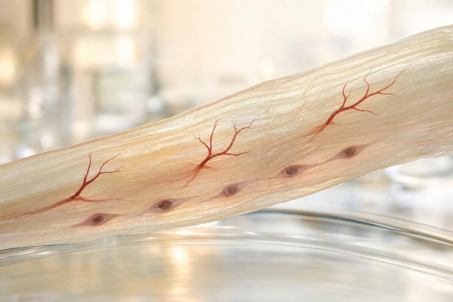
Here’s the short answer: BPC-157 shows repeat lab signals in tendon and ligament injury models, but the data are still almost all from rats, tendon explants, and cell studies. There are no tendon-specific human randomized trials, no FDA-approved use, and no clear human dose for tendon care as of August 17, 2026.
If you want the plain takeaway, it’s this:
- Most data come from rat Achilles tendon studies
- The same lab patterns show up most often in:
- VEGFR2 / Akt / eNOS angiogenesis
- nitric oxide (NO) signaling
- FAK / paxillin fibroblast migration
- collagen fiber organization
- Growth hormone receptor upregulation shows up in some tendon fibroblast work, but less often
- Non-Achilles models like rotator cuff, MCL, quadriceps tendon, and myotendinous junction mostly point in the same direction, but direct pathway testing is thinner
- Reported animal findings include about a 3.8-fold increase in VEGFR2 in one tendon setting and about 30% better maximal tensile strength at 28 days in some rat work
- That does not mean the same effect is proven in people
What I take from this article is simple: the mechanism story is strongest in Achilles models, less direct in other tendon sites, and still far from clinical proof. You can use this research as lab background, but not as proof of human safety, human dosing, or human efficacy.
BPC 157 for tendon and ligament injury healing
sbb-itb-7164bd9
Quick comparison
| Model | Main finding | Mechanism support | What to keep in mind |
|---|---|---|---|
| Achilles tendon | Better load to failure, stiffness, AFI, collagen alignment | Strongest direct data | Still animal-only |
| Rotator cuff | Better function and tendon-to-bone healing | Mostly inferred from Achilles/cell work | Few direct pathway assays |
| MCL | Better healing, earlier vascularization, collagen I/III changes | Some direct vascular data | Ligament data, not tendon mid-substance |
| Quadriceps / MTJ | Reattachment and restored junction structure | eNOS and COX-2 reported directly | Sparse junction-level signaling detail |
So if you’re reading this to answer, “What does BPC-157 seem to do in tendon models?” the plain answer is: it is linked with blood vessel signaling, cell movement, and matrix repair in preclinical studies - mainly in rats. If you’re asking, “Does that prove it works for human tendon injuries?” the answer is no.
That’s the frame I’d use for the rest of the article.
Mechanisms reported repeatedly across tendon and ligament models
Looking across the repeat findings, three themes show up again and again in tendon models: angiogenesis, cell migration, and matrix repair. That said, the evidence base is still early, requiring a clinical overview of peptide therapy to contextualize these findings. A 2025 orthopedic review identified 36 studies, with 35 of them preclinical, which means these findings come from animal and in vitro work, not confirmed human pharmacodynamic data.
Angiogenesis and nitric oxide signaling
The clearest repeat signal is pro-angiogenesis through the VEGFR2-Akt-eNOS pathway, which appears to increase nitric oxide (NO) and support microvessel formation. This pattern shows up most often in Achilles models. It has also been reported in rotator cuff tendon-to-bone repair, though the signal is thinner in ligament models.
In rodent Achilles transection studies, BPC-157 increased vessel density and tensile strength at 14 days compared with saline. When researchers used L-NAME, that effect dropped off, which points to nitric oxide dependence. Some papers also suggest bidirectional NO modulation: boosting eNOS in vascular endothelium while dialing down overactive inducible NOS in inflammatory settings.
After blood supply, the next repeat pattern moves to cell behavior and matrix remodeling.
FAK-paxillin signaling, fibroblast migration, and matrix repair
FAK-paxillin signaling is tied to adhesion, migration, and load signaling. In ex vivo and in vitro tendon models, studies report greater fibroblast outgrowth, migration, and stress resistance, which fits the idea of repair-site repopulation.
In tendon-to-bone healing models, FAK-paxillin activation is proposed to help fibroblasts move back into the repair site and help coordinate extracellular matrix remodeling, including better collagen fiber alignment. Several animal studies report both findings.
Collagen organization, growth hormone receptor pathways, and inflammatory modulation
Other reported effects look more like support players than primary drivers of healing. Three signals come up with some regularity, but not as often.
Type I collagen deposition and better fiber organization appear across several tendon models and are usually read as signs of more mature repair tissue. In some Achilles rupture-repair studies, collagen staining and fiber arrangement improved even when composite histologic scores did not move much.
Growth hormone receptor (GHR) upregulation is reported less often. A 2014 tendon fibroblast study found a 7-fold increase in growth hormone receptor expression, along with dose- and time-dependent increases in proliferation and migration. That finding is worth noting, but it should not be given the same weight as the angiogenesis or FAK-paxillin data.
Anti-inflammatory effects are also mentioned often, including drops in COX-2, MPO, TNF-α, IL-6, and IFN-γ. But these cytokine findings vary from study to study and are less tendon-specific than the angiogenesis and migration signals.
| Mechanism | How Often Reported | Key Signals | Evidence Source |
|---|---|---|---|
| Angiogenesis / NO signaling | Most consistently reported | VEGFR2, Akt-eNOS, NO production | Animal and in vitro tendon/soft-tissue models |
| FAK-paxillin / fibroblast migration | Most consistently reported | FAK, paxillin, cytoskeletal organization | Tendon explants, in vitro studies, animal models |
| Collagen / ECM remodeling | Frequently reported | Type I collagen, fiber alignment | Animal histology and biomechanical testing |
| Inflammatory modulation | Reported across multiple models, but less tendon-specific | COX-2, MPO, TNF-α, IL-6, IFN-γ | Often extrapolated from broader musculoskeletal models |
| Growth hormone receptor signaling | Occasionally reported | GHR gene expression in tendon fibroblasts | Limited tendon-specific studies |
Achilles tendon models: where most mechanistic data come from
Most of the mechanistic detail comes from rat Achilles models. That matters because these studies usually track several things in the same experiment: functional scores, biomechanical testing, histology, and molecular signals. Put simply, this is where the BPC-157 tendon literature is most fully mapped out.
Rat studies usually use one of a few injury setups: mid-substance transection, calcaneal detachment, or tendon-to-bone transection followed by reattachment. Because of that, the Achilles literature gives the clearest test of the angiogenesis, FAK-paxillin, and matrix-repair signals discussed earlier.
Functional, biomechanical, and histologic findings in Achilles studies
The functional outcome reported most often is the rat Achilles Functional Index (AFI), which is a gait-based score. Across studies, BPC-157-treated animals tend to score better than saline controls, with the biggest early gains showing up in the first 4 days in tendon-to-bone detachment models.
Biomechanical results point in the same direction. Treated tendons show higher load to failure, along with higher Young's modulus and stiffness. Reported doses span a preclinical range from 10 pg/kg to 10 µg/kg, given intraperitoneally.
Histology adds more detail. Compared with controls, treated tendons show:
- Smaller defect size
- Better collagen type I fiber alignment
- More organized fibroblast arrays
- A cell pattern that shifts away from acute inflammation, with fewer granulocytes and more mononuclear cells
Early healing phases in detachment models also show increased neovascular indices and more mature neovascularity.
Mechanistic signals observed in Achilles healing
Achilles studies are useful because they test whether broad mechanism claims line up with actual tissue repair. In Achilles explants, BPC-157 increased fibroblast outgrowth and migration, with dose-dependent FAK and paxillin phosphorylation, while total protein expression stayed unchanged. That fits with the better collagen organization and defect closure seen in the in vivo work.
For angiogenesis-related signals, Achilles studies mostly describe vessel changes in vivo rather than direct angiogenesis in isolated culture. Injured tendon tissue has shown higher vessel density and higher VEGF expression after treatment.
Corticosteroid-impaired healing models add another piece. When healing is worsened with methylprednisolone (5 mg/kg), BPC-157 at 10 µg/kg i.p. partly offsets the damage: AFI improves sooner, MPO activity drops, inflammatory cell influx falls, and the neovascular index rebounds.
Achilles study designs, endpoints, and mechanism claims: a comparison table
The table below shows how Achilles BPC-157 studies differ in design, and what those differences mean when reading the mechanism claims.
| Injury Type | Route / Dose | Primary Outcomes | Mechanistic Readouts | Key Limitation |
|---|---|---|---|---|
| Mid-substance transection (rat) | i.p., 10 µg/kg–10 pg/kg daily | Load to failure, Young's modulus, AFI, defect closure | FAK-paxillin activation, VEGF | No human PK/PD data |
| Tendon-to-bone detachment (rat) | i.p., 10 µg/kg daily | AFI (days 1–21), stiffness, neovascular index | Angiogenesis markers, MPO activity, collagen type I | AFI is rat-specific |
| Corticosteroid-impaired detachment (rat) | i.p. BPC-157 + methylprednisolone 5 mg/kg | AFI recovery, MPO, neovascular index | Inflammatory modulation, vascular normalization | Mechanism claims remain correlative |
| Ex vivo tendon explants / fibroblast culture | Direct BPC-157 exposure in vitro | Fibroblast outgrowth, migration, survival | FAK-paxillin phosphorylation | In vitro findings require in vivo confirmation |
The next step is to see whether this same pattern shows up in rotator cuff models and other non-Achilles tendon studies.
Rotator cuff and other models: where findings hold and where they are thin
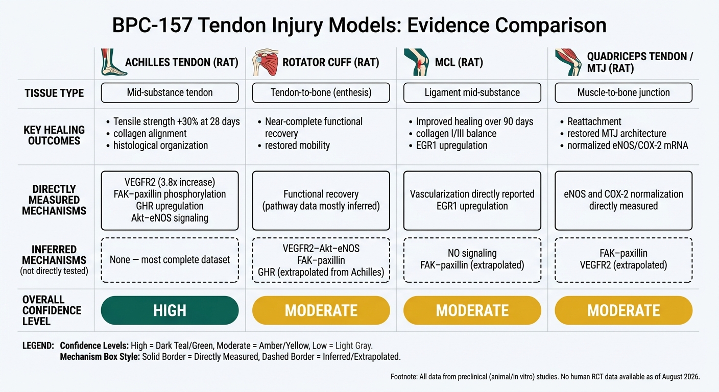
BPC-157 in Tendon Models: Evidence Strength by Injury Site
Rotator cuff tendon-to-bone healing
Rat rotator cuff models, using surgical detachment of the supraspinatus and infraspinatus tendons, report near-complete functional recovery with daily intraperitoneal BPC-157 at 10 µg/kg. Treated animals regained mobility, muscle strength, and leg length measures that came close to uninjured controls. Researchers also noted visible tendon healing, which points to support for tendon-to-bone repair and gross function.
That point matters because healing at the enthesis is not the same as healing in the middle of a tendon. It’s a different biological setting. And here’s where the evidence starts to thin out a bit: biomechanical testing in rotator cuff studies is far less detailed than in Achilles work. These papers rarely include formal load-to-failure curves or stiffness data, and enthesis-level signaling assays are limited.
So the main signal is functional recovery. The mechanism story is less direct. Claims around VEGFR2–Akt–eNOS angiogenesis, FAK–paxillin fibroblast migration, and growth hormone receptor upregulation mostly come from Achilles studies and fibroblast culture work, not from direct measurement in rotator cuff tissue. Put simply, the recovery data are there; the pathway data are mostly inferred.
MCL, quadriceps tendon, and myotendinous junction models
MCL transection studies in rats show better ligament healing for as long as 90 days, along with earlier vascularization and a more favorable collagen type I/III balance. BPC-157 showed effects when given intraperitoneally, orally, or topically as a 1 µg/g cream. Researchers have also reported upregulation of EGR1 (early growth response factor-1), a transcription factor linked to cell growth and inflammation, in MCL tissue.
That lines up well with the angiogenesis and matrix-repair themes seen in Achilles studies. Still, tissue-specific FAK–paxillin measurements in MCL are limited, so those pathway claims still lean heavily on tendon explant and Achilles data.
Quadriceps tendon and myotendinous junction (MTJ) models show much the same pattern. Oral BPC-157 at 10 µg/kg/day and 10 ng/kg/day led to muscle-to-bone reattachment and restored MTJ architecture after surgical detachment of the rectus and vastus muscles. By days 28–42, treated rats moved from large defects with inflammatory infiltrates to a fully re-established MTJ with well-oriented dense connective tissue and no inflammatory cells. Controls, by contrast, still showed defects and atrophy. Studies also reported normalized eNOS and COX-2 mRNA levels, which fits with nitric oxide pathway modulation during recovery.
Alignment and conflict across non-Achilles models: a comparison table
The table below shows where non-Achilles results match Achilles data and where the evidence is lighter.
| Model | Tissue | Main Healing Outcomes | Measured Mechanisms | Confidence Level |
|---|---|---|---|---|
| Achilles (rat) | Tendon mid-substance | Tensile strength, histological organization, collagen alignment | GHR upregulation, FAK–paxillin phosphorylation, VEGFR2–Akt–eNOS signaling | High - directly measured |
| Rotator cuff (rat) | Tendon-to-bone (enthesis) | Near-complete functional recovery, tendon healing, restored mobility and strength | Angiogenesis and matrix repair mostly inferred from Achilles/fibroblast data | Moderate - outcome data present; pathway claims extrapolated |
| MCL (rat) | Ligament mid-substance | Improved healing over 90 days, collagen I/III balance, EGR1 upregulation | Vascularization reported directly; NO/FAK–paxillin mostly inferred | Moderate - consistent with tendon themes; ligament-specific assays limited |
| Quadriceps tendon / MTJ (rat) | Muscle-to-bone junction | Reattachment, restored MTJ architecture, normalized eNOS/COX-2 mRNA | eNOS and COX-2 normalization direct; FAK–paxillin and VEGFR2 inferred | Moderate - functional data strong; junction-specific signaling sparse |
Across these non-Achilles models, the pattern is pretty steady: better healing, improved collagen organization, and earlier vascularization keep showing up. What shows up less often is direct pathway measurement in the exact tissue being tested. Most of the mechanistic detail still comes from Achilles and in vitro fibroblast work, then gets carried over by analogy to rotator cuff, ligament, and junction models.
Clinical interpretation for U.S. clinicians: what the data support and what they do not
What clinicians can reasonably take from the literature
The preclinical BPC-157 tendon literature shows a repeatable pattern. Across studies, the same signals keep showing up: angiogenesis/NO modulation, FAK–paxillin-driven cell migration, and better matrix organization. Separate in vitro work also reports growth hormone receptor upregulation in tendon fibroblasts.
That pattern shows up most clearly in Achilles models. Those studies give the cleanest mechanistic picture because they measured pathway shifts and healing outcomes side by side. In rat transection models, investigators reported a 3.8-fold increase in VEGFR2 expression versus controls and about a 30% improvement in maximal tensile strength at 28 days. Non-Achilles models generally point the same way, but they include less direct pathway data.
For U.S. clinicians, the plain takeaway is this: BPC-157 is still a preclinical candidate, not a proven therapy.
What clinicians should not infer from these studies
Animal and ex vivo findings do not tell us human dosing, human efficacy, or human safety. There are no completed Phase 2 or Phase 3 tendon trials. One Phase I study has been listed since 2016. So there is no validated dose-response curve, no confirmed pharmacokinetic profile, and no support for claiming it works better than standard care such as PRP, rehab, or surgery.
The gap between preclinical promise and human care is still large. Clinical dosing, safety, and regulatory use remain unresolved. Human safety data for musculoskeletal use is basically absent, and much of the literature comes from a small group of labs. On top of that, current FDA policy blocks 503A/503B compounding for human drugs.
Conclusion: the main pattern across tendon injury models
Taken together, the model comparison supports a narrow reading of the evidence. The same preclinical themes show up across tendon injury models, but the data support more study, not clinical adoption.
FAQs
Why are Achilles studies considered the strongest evidence?
Achilles tendon studies are often treated as the strongest support for BPC-157. Why? Because this is the most consistent and best-documented part of the preclinical research.
In rat Achilles tendon transection models, researchers repeatedly report two things:
- Better tendon strength
- Better histological organization
That said, this evidence is entirely preclinical. Results in animals do not guarantee the same results in humans, so they need to be read with caution.
Which BPC-157 mechanisms show up most consistently in tendon models?
The mechanisms reported most often in tendon injury models center on angiogenic growth factors, the nitric oxide (NO) system, and cell signaling tied to tissue repair.
Across these studies, the main patterns are pretty consistent: VEGF upregulation, activation of the Akt-eNOS axis, and the BPC-157–FBXO22–BACH1 pathway. These mechanisms are linked to new blood vessel growth, a return toward normal NO balance, and support for collagen deposition and fibroblast migration.
What can clinicians conclude from these animal studies?
Clinicians should read the evidence on peptides like BPC-157 and TB-500 with caution. At this point, the support behind them comes only from preclinical work in animals and in vitro settings. There are no randomized, controlled human trials that confirm either efficacy or long-term safety.
Animal studies do show positive signals for musculoskeletal repair and wound healing. But that’s not the same as proof in people. A result in a lab or animal model doesn’t guarantee the same outcome in human patients.
For now, these agents are best treated as investigational. That means using thorough informed consent, keeping careful documentation, and paying close attention to contraindications such as malignancy.