RgIA requires about 300 times the concentration to inhibit human α9α10 receptors as rat receptors in the reported assays. I compare receptor species, subunits, and test conditions before drawing conclusions - and you should, too. A single α9 residue, Thr56 in rats versus Ile56 in humans, explains much of that gap.
Here’s how the four peptides compare:
Quick Comparison
| Peptide | Human–rodent comparison | Main limit |
|---|---|---|
| RgIA | IC₅₀: about 490 nM in humans vs. 1.5 nM in rats | Residue 56 drives much of the difference |
| Vc1.1 | About 400-fold weaker inhibition in humans than rats | Receptor blockade and proposed pain mechanisms need separate assessment |
| PeIA | Responses depend on species, receptor setup, and assay | Also inhibits other nicotinic receptors |
| RgIA4 | IC₅₀: about 1.5 nM in humans vs. 0.9 nM in rats | Similar receptor responses do not establish human pain relief |
I keep receptor-current measurements separate from animal pain outcomes. <u>Neither establishes a safe human dose or proves that a treatment works in people.</u> That requires human receptor testing, drug-exposure and safety studies, and clinical trials - not a conversion from a rodent dose.
Human vs. Rodent α9α10 Receptor Pharmacology
Human α9α10 Receptor Structure and Function
Human α9α10 receptors are ligand-gated ion channels made from α9 and α10 subunits. Acetylcholine activates these channels, while nicotine blocks or antagonizes them. Studies have not established a physiologically relevant standalone mammalian α10 receptor. Researchers therefore need to state whether they tested α9 alone or the α9α10 heteromer.
α9α10 receptors have an established role in cochlear hair-cell signaling. But finding receptor expression in immune or non-neural tissues does not prove that functional channels assemble or that targeting them offers clinical benefit. Any immune effect remains experimental. Expression alone also does not establish a shared peptide target across species.
The sequence context of α9 and α10 differs across species, so peptide potency is species-specific - not universal. Rodent peptide data can inform human research, but the results do not transfer directly.
Rat and Mouse α9α10 Receptors in Pain Studies
Rodent models can show whether a peptide alters α9α10-linked phenotypes. They cannot establish its potency at human receptors.
Rat evidence: In rats with chronic constriction injury, RgIA improved pain behavior and tissue findings. These endpoints, however, do not directly measure receptor blockade.
Mouse evidence: RgIA4 prevented oxaliplatin-induced cold allodynia in wild-type mice. In α9-knockout mice, it did not relieve residual allodynia. This supports an α9-dependent component, but it does not prove that every peptide effect acts through that pathway.
The table separates receptor assays from animal outcomes.
| Receptor species | Receptor subunits | Experimental system | Assay format | Peptide | Concentration or dose | Key finding |
|---|---|---|---|---|---|---|
| Human α9α10 | α9 + α10 | Recombinant expression | Receptor-current electrophysiology | RgIA4 | Not provided here | Engineered for high human potency; exact IC50 not provided here. |
| Rat α9α10 | α9 + α10 | Recombinant expression | Receptor-current electrophysiology | Vc1.1, RgIA | Not provided here | Strong inhibition; exact IC50 values not provided here. |
| Mouse α9α10 | α9 + α10 | Recombinant expression | Receptor-current electrophysiology | RgIA4 | Not provided here | Low-nanomolar potency; exact IC50 not provided here. |
| Rat | Not directly reported | Chronic constriction injury | Stimulus-response and weight-bearing tests | RgIA | Not provided here | Improved pain behavior and tissue findings. |
| Wild-type mouse | α9-containing system | Oxaliplatin model | Cold-allodynia testing | RgIA4 | Not provided here | Prevented cold allodynia. |
| α9-knockout mouse | α9 absent | Oxaliplatin model | Cold-allodynia testing | RgIA4 | Not provided here | Did not relieve residual allodynia. |
Before comparing potency, check the subunit ratio, agonist concentration, peptide exposure time, recording conditions, and reversibility. IC50 values and animal doses answer different questions and should not be treated as equivalent.
sbb-itb-7164bd9
RgIA: Human–Rat Inhibition and Sequence Differences
RgIA Potency: Human vs. Rat α9α10 Receptors
RgIA Inhibition of Human, Rat, and Mixed Receptors
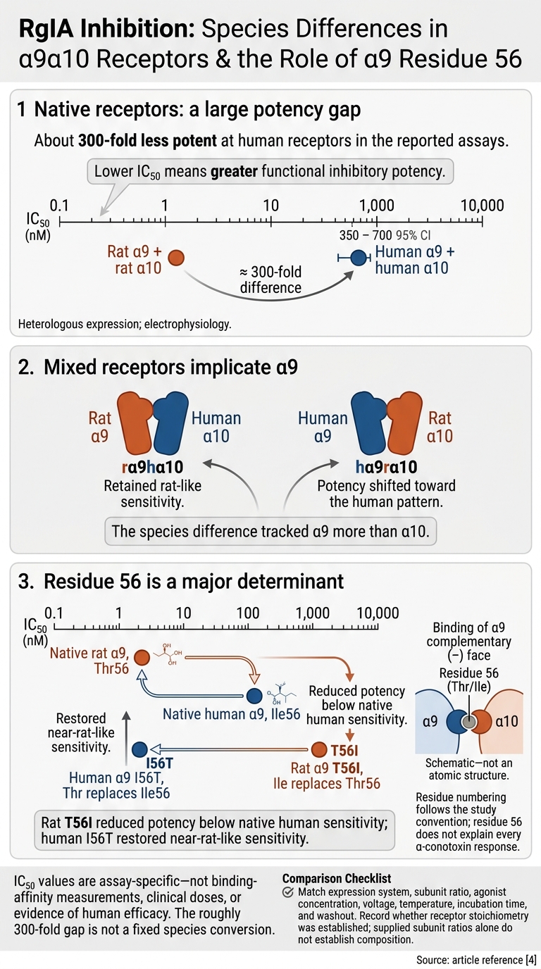
RgIA was about 300-fold less potent at human α9α10 receptors than at rat α9α10 receptors. Tests with mixed receptors showed that the difference tracked α9 more than α10.
| Species or combination | Receptor composition | Assay type | RgIA inhibition metric |
|---|---|---|---|
| Rat | Rat α9 + rat α10 | Heterologous expression; electrophysiology | IC₅₀ ≈ 1.5 nM |
| Human | Human α9 + human α10 | Heterologous expression; electrophysiology | IC₅₀ ≈ 490 nM; 95% CI: 350–700 nM |
| Rat α9 + human α10 | rα9hα10 | Mixed-subunit electrophysiology | Retained rat-like sensitivity |
| Human α9 + rat α10 | hα9rα10 | Mixed-subunit electrophysiology | Potency shifted toward the human pattern |
IC₅₀ values are assay-specific.
This pattern points to residue 56 in α9 as a major driver of the difference.
Rat α9 Thr56 vs. Human α9 Ile56
Rat α9 Thr56 and human α9 Ile56 sit on the complementary (−) face of the binding interface. Swapping these residues in both species showed that this site accounts for much of the species gap. Residue numbers follow the study’s numbering convention.
| Subunit or construct | Residue identity | RgIA IC₅₀ |
|---|---|---|
| Native rat α9 | Thr56 | ≈ 1.5 nM |
| Native human α9 | Ile56 | ≈ 490 nM |
| Rat α9 T56I | Ile replaces Thr56 | ≈ 2.5 µM |
| Human α9 I56T | Thr replaces Ile56 | ≈ 1.9 nM |
Rat T56I reduced RgIA potency below that seen with native human α9α10. Human I56T, meanwhile, restored near-rat-like sensitivity. Residue 56 is a major determinant for RgIA, but it does not explain every α-conotoxin response.
Separating Sequence Effects From Assay Conditions
Potency differences also depend on how the assay is run. For comparisons, match the expression system, subunit ratio, agonist concentration, voltage, temperature, incubation time, and washout. The roughly 300-fold gap came from specific assay conditions; it is not a fixed conversion between species.
Also record whether receptor stoichiometry was established. The supplied subunit ratio alone does not prove the receptor’s subunit composition.
IC₅₀ values are not clinical dose or efficacy estimates, and sequence similarity does not justify converting a rodent dose into a human dose.
Vc1.1, PeIA, and RgIA4: Cross-Species Results
Beyond RgIA, these peptides differ in potency across species, receptor selectivity, and engineered cross-species activity. The table keeps functional potency, binding affinity, selectivity, and animal outcomes separate. “Not reported” means no result was provided - not that activity is absent. Values from different assays should not be ranked against one another.
| Peptide | Receptor species | Experimental system | Functional potency | Binding affinity | Selectivity | Animal outcomes |
|---|---|---|---|---|---|---|
| Vc1.1 | Human vs. rat α9α10 | Recombinant receptor assays; neuronal calcium-current studies | ≈400-fold less potent at human α9α10 than rat | Not directly measured | α9α10 antagonism; separate studies also report GABA_B-linked N-type calcium-channel inhibition | Reduced tactile allodynia and mechanical hyperalgesia in rodent neuropathic-pain models |
| PeIA | Rat α9α10 and selected nAChR subtypes; no matched human–mouse comparison reported | Recombinant nAChR subtype assays; rat dorsal-root-ganglion neuron recordings | α9α10 IC₅₀ ≈ 30 nM; neuronal calcium-current IC₅₀ ≈ 1.1 nM at a different endpoint | Not directly measured | Incomplete selectivity: also inhibits α3β2 and α6/α3β2β3 at ~20 nM | No specific animal analgesic outcome established in the cited sources |
| RgIA4 | Human vs. rat α9α10; no mouse functional value reported | Recombinant human and rodent receptor assays; rat and mouse pain models | Rat IC₅₀ ≈ 0.9 nM; human IC₅₀ ≈ 1.5 nM | Not directly measured | Reported >1,000-fold selectivity over tested off-targets, including opioid and GABA_B receptors | Prevented oxaliplatin-induced pain behaviors in rats and mice |
Vc1.1 and PeIA: Receptor Sensitivity and Selectivity
Vc1.1 and PeIA have different limits: Vc1.1’s potency varies by species, while PeIA acts on receptors beyond α9α10.
For Vc1.1, the human–rat difference reflects functional inhibition, not a directly measured binding constant. Its proposed pain-relief mechanisms also need to stay separate: α9α10 blockade and GABA_B-linked calcium-channel inhibition are distinct findings.
PeIA is better described as a tool for distinguishing receptor subtypes than as an α9α10-specific peptide. Its activity at other nicotinic receptors rules out that narrower label.
RgIA4: Engineering for Human and Rodent Receptors
Unlike Vc1.1 and PeIA, RgIA4 was designed to narrow the human–rodent potency gap. This synthetic, second-generation RgIA analog addresses weak activity at the human receptor. The paired rat and human measurements support improved cross-species functional potency - not directly measured binding affinity.
The rodent pain results are separate preclinical evidence. When recording these findings, keep the species, chemotherapy model, behavioral endpoint, and available dosing details. Mark missing fields rather than filling them in by inference.
Conclusion: What Receptor Studies Cannot Establish
Across the peptides reviewed above, sequence differences can change inhibition. But recombinant current block and rodent behavior do not establish human efficacy or prove that α9α10 blockade alone caused an effect.
Evidence Needed Before Human Dosing
Human dosing must rest on direct translational data. That means testing human receptors, using matched rodent assays to separate sequence effects from assay conditions, and conducting species-appropriate pharmacokinetic and safety studies. These studies must address tissue distribution, metabolism, clearance, and off-target activity.
Validated exposure–response models should link unbound peptide concentrations to receptor occupancy, pharmacodynamic effects, and adverse effects. Before making efficacy claims, researchers must conduct dose-escalation studies with pharmacokinetic sampling and prespecified safety monitoring. Randomized controlled trials must then show meaningful pain relief and improved function.
Recording Translation Limits in Clinical Resources
Clinical resources must state these limits clearly. PeptidePrescriber is an educational resource for licensed healthcare professionals. It does not establish human dosing safety or support treating patients with research α-conotoxins.
Monographs should document receptor species, α-subunit composition, expression system, assay conditions, inhibition endpoint, peptide concentration units, sequence variants, and whether each study used animal, recombinant, or human data. Summaries should explicitly state that rodent α9α10 findings alone cannot support a human dosing protocol.
FAQs
How can researchers separate sequence effects from assay differences?
Compare like with like: control for species, receptor system, endpoint, concentration, and exposure duration. Binding affinity, functional potency, and efficacy measure different things and are not interchangeable.
Support receptor attribution with evidence from several approaches: selective pharmacology, functional recordings, and genetic models, such as deletion or rescue. Check for factors that could skew the results, including supraphysiological exposure, inadequate antagonist selectivity, and differences in signaling cascades within the assay system.
Does RgIA4’s human receptor potency predict pain relief?
No. Rodent findings on RgIA4 potency at the α9α10 receptor cannot establish human dosing or efficacy - or predict pain relief in patients.
Receptor sequences, binding, and downstream signaling can differ between species. Animal data therefore help form hypotheses; they don't prove clinical effects in humans. Clinicians need human-specific pharmacokinetic and pharmacodynamic data to determine therapeutic efficacy and safety.
What evidence is needed to establish a safe human dose?
Randomized, controlled human trials offer the strongest evidence for a safe human dose. Because human and animal physiology differ, animal findings cannot reliably establish dosing or efficacy in humans.
When human data are limited, avoid simple weight-based allometric scaling - it can lead to dosing errors. Instead, use condition-specific evidence, monitor safety, start low and titrate slowly, and document clinical responses at every stage.