
Oxytocin and vasopressin differ by just two amino acids, but their receptor effects aren’t interchangeable. My starting point: check the receptor, tissue, species, and exposure before applying a clinical overview of peptide therapy to patient care.
Here, cross-talk means one peptide activates the other’s receptors - not that the receptors interact directly. I focus on three questions:
- Which receptor caused the response? Binding, cell signaling, and patient outcomes answer different questions.
- Did the peptide reach that tissue? Blood levels alone don’t prove brain exposure or receptor activation.
- Does human research support clinical use? Route, dose, risks, and monitoring require human data and current U.S. prescribing information.
<u>Receptor cross-talk alone does not establish safety or effectiveness.</u> I use that distinction to separate what laboratory studies suggest from what clinicians can apply.
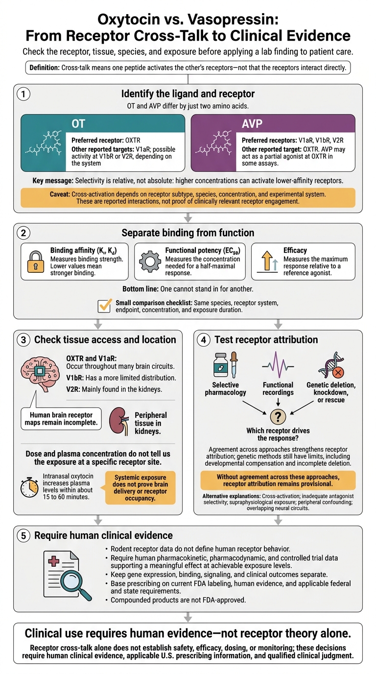
Oxytocin vs. Vasopressin: From Receptor Cross-Talk to Clinical Evidence
Ligand Selectivity and Receptor Cross-Activation
OT and AVP differ by only two amino acids, so each can bind some of the other’s receptors, usually with a lower preference. Selectivity is relative, not absolute: higher concentrations can activate lower-affinity receptors. The key question is which receptor drives the observed effect.
Preferred Receptors and Other Targets
Cross-activation depends on the receptor subtype. AVP can activate OXTR and may act as a partial agonist at OXTR in some assays. OT activity at vasopressin receptors depends on the subtype, species, concentration, and experimental system.
| Ligand | Preferred receptor(s) | Other reported targets | Relative selectivity | Interpretation limits |
|---|---|---|---|---|
| OT | OXTR | V1aR; possible activity at V1bR or V2R, depending on the system | Generally favors OXTR, but selectivity is relative | Depends on the assay system and exposure conditions. |
| AVP | V1aR, V1bR, V2R | OXTR | Some binding studies show similar affinity across OXTR, V1aR, and V1bR | Binding does not establish functional relevance. |
Binding Affinity and Functional Response
Cross-activation matters only if the measured effect occurs at exposures relevant to humans. Three measures describe different parts of that response:
- Binding affinity (Ki, Kd) measures binding strength. Lower values mean stronger binding.
- Functional potency (EC50) measures the concentration needed for a half-maximal response.
- Efficacy measures the maximum response relative to a reference agonist.
These measures answer different questions. One cannot stand in for another. For practitioners, applying these metrics requires following practical clinical guides to ensure precise administration.
Receptor density and receptor reserve can amplify the effects of weak agonists. Exposure duration, peptide breakdown, and the measured endpoint also affect the results. Calcium release, cyclic AMP, and tissue contraction assays may rank the same ligands differently.
Compare like with like: the same species, receptor system, endpoint, concentration, and exposure duration. A detectable laboratory response does not establish a clinically relevant interaction. Receptor location and tissue access determine whether cross-activation can matter in vivo.
sbb-itb-7164bd9
Receptor Locations and Biological Effects
Central and Peripheral Receptor Locations
A receptor’s location tells us where a response could occur - not which receptor caused it. OXTR and V1aR occur throughout many brain circuits. V1bR has a more limited distribution, while V2R is mainly found in the kidneys. Maps of these receptors in the human brain remain incomplete.
Cross-talk matters most where receptor locations overlap and peptides reach those sites. That overlap helps explain why OT and AVP can produce similar effects in some tissues and different effects in others.
| Receptor | Canonical ligand | Central locations | Peripheral locations and effects | Typical signaling |
|---|---|---|---|---|
| OXTR | OT | Hypothalamus, limbic system, hippocampus, and basal forebrain; varies by species | Uterus and mammary tissue: contraction and milk ejection | Usually Gq/11 → PLC → intracellular calcium; other pathways occur |
| V1aR / AVPR1A | AVP | Hypothalamus, lateral septum, and amygdala, among other regions; varies by species | Vascular smooth muscle: vasoconstriction | Usually Gq/11 → PLC → calcium |
| V1bR / AVPR1B | AVP | Selected regions, including hippocampal circuits studied in social memory | Anterior pituitary corticotrophs: ACTH release | Usually Gq/11-linked |
| V2R / AVPR2 | AVP | Much less prominent centrally | Renal collecting ducts: water reabsorption | Primarily Gs → adenylyl cyclase → cAMP → PKA |
Receptor coupling varies with cell type, ligand concentration, receptor density, and conditions. Physiologic state can also change receptor availability and signaling. Human receptor mapping depends on tissue quality, assay sensitivity, and validated receptor-selective ligands.
Keep gene expression, binding, signaling, and clinical outcomes separate. Receptor distribution alone cannot identify which receptor drives an effect in a patient. Likewise, a kidney response does not establish a central effect.
Peptide Exposure, Clearance, and Receptor Regulation
Endogenous peptide release can be synaptic, dendritic, paracrine, or neurosecretory. Clearance and peptide-degrading enzymes - including oxytocinase and vasopressinase - limit exposure.
Dose and plasma concentration do not tell us the exposure at a specific receptor site. Central and peripheral exposure need separate assessment. From there, the question is which receptor best explains the observed response.
Identifying Which Receptor Drives a Response
A positive assay result alone does not establish receptor involvement. Record the ligand, dose or concentration, route, timing, species, sex, age, physiologic state, tissue, receptor-assay method, antagonist or agonist selectivity, endpoint, and the authors’ receptor attribution. Also assess other explanations: cross-activation, inadequate antagonist selectivity, supraphysiological exposure, peripheral confounding, and overlapping neural circuits.
Evidence from several approaches - selective pharmacology, functional recordings, and genetic deletion, knockdown, or rescue - can strengthen receptor attribution when the findings agree. Genetic methods still have limits, including developmental compensation and incomplete deletion.
Without agreement across these approaches, receptor attribution remains provisional. In behavioral studies, describe a response as consistent with receptor involvement unless convergent evidence supports receptor mediation. A laboratory response does not prove patient benefit. Clinicians should consult prescribing guidance to evaluate how these mechanisms translate to practice.
Limits of Translation to U.S. Clinical Practice
Species Differences and Research-Tool Limits
Receptor cross-activation in the lab may not carry over to human biology. Rodent receptor data do not define human receptor behavior. Receptor affinity, distribution, and signaling can differ across species. Radioligands and antagonists may also lose selectivity outside their validated concentration range. Reported selectivity differences range from roughly 10- to 100-fold, depending on the assay and species.
Treat receptor mRNA as supporting evidence - not proof of surface protein or function. Look for validated protein detection, binding, and signaling in relevant human tissue.
Human Exposure and Clinical Outcomes
Intranasal oxytocin increases plasma levels within about 15 to 60 minutes. That shows systemic exposure, but it does not prove brain delivery or receptor occupancy. Exposure depends on clearance, breakdown, formulation, and route. Higher concentrations can reduce receptor selectivity, and behavioral effects may still come from peripheral or indirect actions.
Apply a finding only when human pharmacokinetic, pharmacodynamic, and controlled trial data support a meaningful effect at achievable exposure levels. Age, sex, pregnancy, stress, kidney function, and disease can alter exposure and response. Do not assume results from healthy, nonpregnant adults apply to pregnant patients or people with cardiovascular or renal disease.
Prescribing Limits and Documentation
If human exposure and outcomes do not support the proposed mechanism, prescribing must follow labeling and documented evidence - not receptor theory. Base prescribing on current FDA labeling, human evidence, and applicable federal and state requirements.
Keep labeled uses, such as oxytocin for labor induction, separate from off-label uses of approved products and investigational uses that require appropriate oversight. Compounded products are not FDA-approved. Document the product and formulation, indication, evidence basis, material risks discussed, measurable endpoint, monitoring, and stopping criteria.
Oxytocin monitoring must address uterine hyperstimulation and fluid-related hyponatremia. Vasopressin monitoring must address cardiovascular effects, hyponatremia, and ischemia.
Conclusion: Key Points for Clinical Interpretation
The mechanistic data call for caution: selectivity is relative, not absolute. Cross-talk depends on receptor subtype, concentration, tissue, species, and assay system. Binding affinity and functional response aren't interchangeable, and neither alone shows what drives a response.
When reporting findings, use “may activate” rather than “targets” unless human evidence supports relevant exposure and receptor engagement. Receptor expression or activation in the lab does not establish clinical benefit or CNS engagement.
Patient selection requires human evidence - not receptor theory alone.
Caution: Receptor cross-talk alone does not establish safety, efficacy, dosing, or monitoring; these decisions require human clinical evidence, applicable U.S. prescribing information, and qualified clinical judgment.
FAQs
Can repeated exposure change receptor selectivity?
Yes. Repeated exposure can reduce effective selectivity through receptor desensitization and downregulation. These processes affect the response in different ways.
Desensitization is fast and reversible: repeated stimulation weakens signaling even while receptors remain present. Downregulation is slower and lasts longer: chronic stimulation can move receptors inside cells or cause them to be broken down. This leaves fewer functional receptors and changes the overall response.
Oxytocin protocols involving chronic use should therefore account for possible desensitization.
How might kidney disease affect receptor cross-talk?
Kidney disease can slow the clearance of oxytocin and vasopressin, leading to higher levels in the bloodstream. This may increase unintended receptor cross-talk, where one peptide activates the other’s receptors. That interaction could increase water retention and the risk of hyponatremia (low blood sodium).
Clinicians should check kidney function and electrolytes, especially serum sodium, at baseline and periodically to account for these changes in peptide clearance.
What human evidence would make cross-talk clinically actionable?
Cross-talk between oxytocin and arginine vasopressin receptors needs testing in well-designed, randomized controlled human trials before it can guide clinical practice. Current evidence comes largely from animal studies and small, preliminary human trials with limited statistical power or inconsistent results.
These trials must show measurable improvements in patient outcomes, not just changes in surrogate markers. Researchers must clearly link those improvements to the intervention and account for the risk of false positives in small, early-phase studies.