One pituitary lab value can mislead you. I read these results by pairing each pituitary hormone with its downstream hormone, then checking timing, pulses, and drug effects before I make sense of the panel.
Here’s the short version:
- GH is pulse-based, so a random GH level is often weak; IGF-1 is usually the better screening marker.
- TSH must be read with free T4; a “normal” TSH can still fit central hypothyroidism if free T4 is low.
- ACTH and cortisol depend on clock time; an 8:00 AM sample means less if sleep, stress, illness, or steroid use changed the axis.
- GnRH works by pulses, so LH and FSH patterns make more sense when I check sex steroids, cycle phase, age, and meds.
- Feedback loops matter: cortisol feeds back on ACTH/CRH, thyroid hormone feeds back on TSH/TRH, and sex steroids plus inhibins feed back on LH/FSH.
- Dynamic testing is often needed when static labs sit in the gray zone:
- GH deficiency: stimulation testing
- GH excess: 75-g OGTT with GH suppression
- Adrenal insufficiency: cosyntropin testing
- Suspected Cushing syndrome: late-night salivary cortisol, 24-hour urinary free cortisol, or 1-mg dexamethasone suppression
A few numbers help frame the workup:
- Morning cortisol below about 5 µg/dL may support adrenal insufficiency in the right setting.
- Morning cortisol around 5 to 10 µg/dL is often indeterminate.
- After a 1-mg overnight dexamethasone test, failure to suppress serum cortisol below about 1.8 µg/dL can support hypercortisolism, depending on the assay.
- For GH excess, some labs use GH suppression cutoffs near 1 ng/mL or 0.4 ng/mL during OGTT, based on assay method.
If I had to boil the whole article down to one rule, it would be this: <u>read the whole axis, not one number</u>. That means hypothalamic signal, pituitary output, target-organ hormone, collection time, and confounders such as glucocorticoids, oral estrogen, opioids, dopamine-blocking drugs, acute illness, obesity, nutrition, and sleep.
This article is a quick clinical map for GHRH, somatostatin, TRH, CRH, and GnRH - with the main lab traps I watch for in endocrine care.
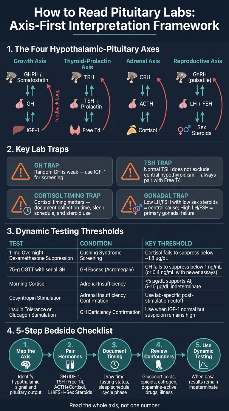
Pituitary Axis Interpretation Framework: Axis-First Clinical Checklist
Endocrinology | Hypothalamus: Anterior Pituitary Connection
sbb-itb-7164bd9
Growth axis control: GHRH and somatostatin
GHRH pushes GH release, and somatostatin puts the brakes on it. That back-and-forth creates pulsatile GH secretion instead of a steady baseline. For clinicians, this is a big deal: GH is one of the hardest pituitary hormones to judge from a single blood draw.
How GHRH stimulates GH and somatostatin suppresses it
The biggest normal GH pulse usually shows up soon after a person falls asleep, especially during slow-wave sleep. After that, GH drives IGF-1 production. IGF-1 lasts longer in circulation, so it reflects GH activity better over time and also helps shut the system back down. GH and IGF-1 both feed back at the pituitary and hypothalamus, lowering GH drive, increasing somatostatin tone, or both.
A lot can tilt this balance. Deep sleep and exercise usually push GH upward. Obesity, hyperglycemia, aging, and glucocorticoid exposure tend to push it downward. Puberty and sex-steroid exposure usually increase GH pulse amplitude. So if an older adult with central obesity has a low GH level, that may fit normal physiologic suppression rather than a pituitary problem on its own.
That’s the key idea here: pattern matters more than one isolated number.
| Signaling state | Expected GH pattern | Expected IGF-1 pattern | Pitfalls |
|---|---|---|---|
| GHRH-dominant | More frequent or higher-amplitude pulses; a random draw may still be low between bursts | Often increased if hepatic and nutritional responsiveness are intact | A high GH pulse alone does not establish GH excess; age, assay method, liver function, and exogenous agents all affect IGF-1 |
| Somatostatin-dominant | Smaller or less frequent pulses; low single GH value may reflect normal timing or physiologic suppression | May be low, but can remain normal in partial or recent suppression | Obesity, aging, hyperglycemia, glucocorticoids, and illness can produce similar findings without permanent GH deficiency |
| Normal pulsatile secretion | Highly time-dependent; rises and falls over minutes to hours | More stable than GH; generally more useful for screening | Interpret with age-, sex-, and assay-adjusted ranges alongside full clinical context |
Before interpreting either GH or IGF-1, factor in age, sex, body composition, pubertal status, medications, nutrition, assay calibration, and the full clinical picture. Miss one of those, and the lab can send you down the wrong path.
Why random GH testing is weak and IGF-1 is more useful
Because GH comes out in short bursts, a random GH value is weak as a screening test. IGF-1 is usually the better integrated marker.
Still, IGF-1 has its own traps. Liver disease, malnutrition, poorly controlled diabetes, hypothyroidism, and oral estrogen exposure can all lower IGF-1 without showing true GH deficiency. In those settings, a low IGF-1 should not be treated as simple proof of GH deficiency. On the flip side, a normal IGF-1 does not exclude GH deficiency, especially in adults with partial deficiency or major comorbid illness.
When the labs and the clinical picture don’t line up, that’s when static testing stops being enough.
When stimulation or suppression testing is needed
Basal labs are for screening. Dynamic testing confirms the diagnosis. For suspected GH deficiency, if clinical suspicion stays high despite a normal IGF-1, move to provocative testing. Validated options include insulin tolerance testing when it is safe and suitable, or glucagon stimulation testing when insulin-induced hypoglycemia is contraindicated or not a good fit.
For suspected GH excess, start with an elevated or equivocal age-adjusted IGF-1. If the answer is still unclear, the next step can be an oral glucose tolerance test (OGTT) with a 75-g glucose load and serial GH measurements. Normal hyperglycemia suppresses GH. If GH fails to suppress below the lab- and guideline-appropriate threshold, that supports autonomous GH excess. Use the lab’s validated suppression cutoff, commonly 1 ng/mL or 0.4 ng/mL with newer assays. Assay calibration differs a lot from one lab to another, so the number only means something when matched to that lab’s reference range.
Dynamic testing can also mislead you if the patient has untreated hypothyroidism, uncontrolled diabetes, acute illness, marked obesity-related suppression, or recent exposure to exogenous GH, GH secretagogues, glucocorticoids, or somatostatin analogs. Those confounders should be documented before testing.
TRH and CRH axes: thyroid-prolactin and adrenal regulation
The best way to read TRH and CRH is to follow the axis all the way through: look at the downstream hormone, check the timing, and ask what feedback signals might be changing the picture.
TRH, TSH, and prolactin: reading the thyroid-prolactin interface
Pituitary results make more sense when you pair the pituitary hormone with the target-organ marker and the clinical picture. TRH stimulates both TSH and prolactin, while dopamine holds prolactin back. Because of that split control, prolactin can go up for more than one reason. It may reflect a pituitary lesion, stalk compression, dopamine-blocking drugs, or severe primary hypothyroidism causing excess TRH.
TSH and free T4 are the key pair for sorting primary from central thyroid disease. A high TSH with a low free T4 points to primary thyroid failure. A low or inappropriately normal TSH with a low free T4 points toward central hypothyroidism, where the pituitary is not responding the way it should. This is where people can get tripped up: central hypothyroidism is easy to miss because TSH may still sit inside the reference range even when free T4 is low.
Severe primary hypothyroidism can also push prolactin up along with TSH. So if a patient has fatigue, menstrual irregularity, a markedly elevated TSH, low free T4, and a modest prolactin elevation, that may be thyroid-driven hyperprolactinemia rather than a prolactinoma. Treat the hypothyroidism first. Then recheck prolactin before calling it a pituitary lesion.
| Clinical pattern | TSH | Free T4 | Practical interpretation |
|---|---|---|---|
| Primary thyroid failure | High | Low | Thyroid failure; prolactin may normalize after treatment. |
| Central hypothyroidism | Low, normal, or inappropriately normal | Low | Pituitary or hypothalamic disease; normal TSH does not exclude. |
| Non-thyroidal illness | Low, normal, or transiently abnormal | Low, normal, or low-normal | Acute illness effect; avoid diagnosing chronic thyroid disease from one panel. |
If prolactin is elevated, start with the basics. Check pregnancy or lactation status, renal function, and every medication the patient is taking. Antipsychotics, antiemetics, antidepressants, and opioids can all contribute. That medication review matters before blaming TRH excess or pituitary disease. Pituitary stalk disruption can also increase prolactin on its own, without TRH being the main driver.
The same axis-based reading applies to cortisol, but here timing and stress can change the result in a big way.
CRH, ACTH, and cortisol: timing and stress effects
The adrenal axis runs as CRH → ACTH → cortisol. Cortisol then feeds back negatively on both the hypothalamus and the pituitary. It normally peaks close to wake time and falls to its lowest point during sleep. That daily rhythm makes collection time one of the first things to document.
An 8:00 AM cortisol after a normal night of sleep is not the same as an 8:00 AM cortisol after night shift work, a hospital stay, or major sleep disruption. Document the exact collection time, sleep-wake schedule, and shift-work status with every result. Acute illness, pain, surgery, stress, and exercise can all increase ACTH and cortisol without meaning the patient has Cushing syndrome.
Exogenous glucocorticoids are another major source of confusion. Oral, inhaled, injected, topical, intra-articular, and even some ophthalmic steroid products can suppress CRH and ACTH and lower endogenous cortisol production. Before reading any adrenal lab, get the full steroid history:
- formulation
- dose
- route
- duration
- time since the last dose
ACTH and cortisol should be read together. That pairing helps sort whether the problem sits in the hypothalamus, pituitary, adrenal gland, or comes from exogenous steroid suppression.
| Clinical pattern | ACTH | Cortisol pattern | Practical interpretation |
|---|---|---|---|
| Primary adrenal insufficiency | High | Low morning cortisol; inadequate cosyntropin response | Adrenal failure; consider mineralocorticoid deficiency. |
| Secondary or tertiary adrenal insufficiency | Low or inappropriately normal | Low morning cortisol | Pituitary/hypothalamic disease or prior glucocorticoid exposure. |
| ACTH-dependent hypercortisolism | High or inappropriately normal | Loss of diurnal nadir and failed suppression | Confirm hypercortisolism before localization. |
| Exogenous glucocorticoid suppression | Low or suppressed | Endogenous cortisol low or inadequate for physiologic needs | Review all steroid routes and duration; assess HPA-axis recovery during supervised tapering. |
When borderline results should not be overcalled
A single borderline TSH or cortisol result usually is not enough to confirm or rule out central dysfunction. On the adrenal side, a morning cortisol below about 5 µg/dL can support adrenal insufficiency in the right setting, but the cutoff depends on the assay. Results in the range of about 5 to 10 µg/dL are often indeterminate and usually need confirmatory testing, most often a cosyntropin stimulation test with cortisol measured before and after synthetic ACTH, using the lab’s assay-specific cutoff.
For suspected Cushing syndrome, random serum cortisol is not a good screening test, especially during acute stress or hospitalization. Accepted first-line screening options include at least two late-night salivary cortisol measurements, at least two 24-hour urinary free cortisol collections, or a 1-mg overnight dexamethasone suppression test. Failure to suppress serum cortisol below about 1.8 µg/dL (50 nmol/L) after the 1-mg test is a common cutoff, but the result still depends on the assay, dexamethasone exposure, adherence, and drug interactions. If results come back abnormal, that should lead to endocrinology review, not a straight jump to imaging.
In day-to-day endocrine work, it helps to lock down the context at the start: collection time, sleep schedule, acute illness, medication exposure, and steroid use. That simple habit can prevent a lot of reading errors.
GnRH follows the same basic rule: pulse pattern, not one isolated value, drives LH and FSH output.
GnRH and reproductive signaling: LH and FSH through pulsatile control
GnRH is released in pulses, and that timing is what keeps gonadotrophs responsive. In plain English: the pituitary does not want a nonstop GnRH signal, a concept central to understanding peptide therapy and its clinical application. It responds best when GnRH arrives in short bursts. When that pulse pattern stays intact, LH and FSH are released in a coordinated way. When pulsatility breaks down, LH and FSH drop.
Pulsatile GnRH versus continuous exposure
GnRH pulse frequency and amplitude help shape LH and FSH output. Broadly speaking, faster pulses tend to push LH more, while slower pulses support proportionally greater FSH release. But this isn't locked in stone. Gonadal steroids, inhibin, activin, sex, age, and menstrual-cycle stage all change how the pituitary responds.
The clinical point is the difference between pulsatile and continuous stimulation. Continuous GnRH receptor activation can cause a brief early rise in LH and FSH. After that, sustained exposure leads to receptor downregulation and pituitary desensitization. Then output falls.
That explains how long-acting GnRH agonists work in practice: they suppress gonadotropins after the initial "flare." So the takeaway is simple: more GnRH does not automatically mean more LH and FSH.
That contrast also helps explain lab patterns used to sort central disease from primary gonadal disease.
Central hypogonadism, primary gonadal failure, and functional suppression
Low testosterone or estradiol with low or inappropriately normal LH and FSH points to central or functional suppression. In that setting, the hypothalamus or pituitary is not providing enough drive. By contrast, low sex steroids with high LH and FSH suggest primary gonadal failure, where the gonads are not responding despite normal or increased upstream drive.
There are a few catches. In men, low inhibin B from Sertoli-cell dysfunction can push FSH up before LH starts to rise. In women, inhibin A and inhibin B shift across the cycle, so one isolated result should not be used to diagnose ovarian failure.
Functional suppression of GnRH pulsatility deserves close attention because it can look exactly like organic central hypogonadism on a standard lab panel. Functional hypothalamic suppression often shows low or low-normal LH, FSH that is not appropriately elevated, low estradiol, and low progesterone. The difference usually comes from the clinical setting. Low energy availability, high training load, restrictive eating, psychological stress, and chronic illness can all disrupt GnRH pulsatility without structural pituitary disease. Research settings have linked energy availability below about 30 kcal/kg of fat-free mass per day with impaired LH pulsatility, and normal body weight does not rule this out.
| Clinical pattern | GnRH drive | LH | FSH | Sex steroids | Interpretation limits |
|---|---|---|---|---|---|
| Hypothalamic or pituitary hypogonadism | Low or ineffective | Low or inappropriately normal | Low or inappropriately normal | Low testosterone or estradiol | A "normal" LH or FSH is inappropriate when sex steroids are low; assess prolactin, other pituitary hormones, and medications |
| Primary gonadal failure | Increased (reduced negative feedback) | Elevated | Elevated; may be especially prominent when inhibin feedback is reduced | Low testosterone or estradiol | Partial failure may produce borderline values; cycle stage, age, and exogenous hormones affect results |
| Functional suppression of GnRH pulsatility | Reduced GnRH pulse frequency or amplitude | Low or low-normal, often lower than FSH | Low-normal or normal, often higher than LH | Low estradiol; testosterone may be low in men | Clinical and exclusionary diagnosis; pregnancy, thyroid disease, hyperprolactinemia, and structural pituitary disease must be assessed |
| Hyperprolactinemia-associated suppression | Functionally suppressed by elevated prolactin | Low or inappropriately normal | Low or inappropriately normal | Low testosterone or estradiol | Confirm persistent elevation; review medications, macroprolactin, and pituitary disease when indicated |
Why reproductive labs must be timed and contextualized
Testosterone, estradiol, LH, and FSH only make sense when you read them with the collection time, sex, age, and cycle phase in mind. In men, testosterone should be drawn in the morning and confirmed if it comes back unexpectedly low before labeling the disorder. LH and FSH then help separate primary testicular dysfunction from secondary hypogonadism. The Endocrine Society recommends checking both in men with confirmed hypogonadism for that reason. In women, estradiol and gonadotropins move quite a bit across the follicular, ovulatory, and luteal phases, so one result without cycle timing is often hard to read.
Medication review comes first. Exogenous testosterone, anabolic-androgenic steroids, opioids, glucocorticoids, hormonal contraception, antipsychotics, and dopamine-modifying drugs can shift the axis enough to mimic disease or hide it. When results are borderline, discordant, or don't fit the clinical picture, repeat testing or dynamic testing usually tells you more than one isolated panel.
Interpret these values alongside the downstream sex steroid result, not as stand-alone numbers.
Clinical integration for peptide-based care and conclusion
To read pituitary function well, you need the full axis, not just one lab on its own. That means looking at the hypothalamic signal, the pituitary hormone, the downstream hormone, the timing of the blood draw, and the feedback loops that shape the result.
A simple bedside approach works best: axis first, then timing, then confounders.
A practical axis-based interpretation checklist
Before acting on any pituitary lab result, work through these steps in order:
- Map the axis - Define the hypothalamic signal and pituitary output: GHRH/somatostatin → GH, TRH → TSH/prolactin, CRH → ACTH, GnRH → LH/FSH.
- Pair pituitary and target-organ hormones - GH with IGF-1, TSH with free T4, ACTH with morning cortisol, LH/FSH with sex steroids.
- Document draw time and major confounders - fasting, sleep, cycle phase, and acute illness.
- Review medications - glucocorticoids, exogenous sex steroids, opioids, dopamine-active drugs, and thyroid hormone can all shift results enough to mimic or mask disease. Review glucocorticoid exposure before HPA-axis testing.
- Use dynamic testing when basal results remain indeterminate.
Once you’ve mapped the axis, tools can help keep dosing, documentation, and lab tracking organized.
Where peptide-focused clinical tools fit in practice
Clinical tools can support the workflow, but they do not replace axis-based interpretation.
For licensed clinicians working in peptide-aware settings, PeptidePrescriber is a clinician resource with peptide monographs, dosing protocols, calculators, injection guides, regulatory references, and pharmacy-agnostic sourcing guidance. In practice, it helps with structured education, documentation, and compliance. It does not replace endocrine evaluation, prescribing standards, or specialist consultation.
Key takeaways for endocrine interpretation
Hypothalamic peptides shape pituitary output through stimulation, inhibition, pulsatility, circadian timing, and feedback. That’s why no single lab value tells the whole story. A pituitary hormone may sit in the “normal” range and still be physiologically inappropriate if the downstream target-organ hormone is low.
A few points matter more than most:
- Random GH is weak.
- IGF-1 is better for screening, but suspected GH deficiency still needs stimulation testing.
- Routine GnRH testing adds little in hypopituitarism, and dynamic TSH testing is not recommended for diagnosing central hypothyroidism.
When results land in the gray zone, the next move is verification and context, not automatic diagnosis or treatment. If multiple axes are off, symptoms point to mass effect, adrenal insufficiency is on the table, or repeat testing still leaves a mismatch, endocrinology referral is the right next step.
This educational framework does not replace specialist consultation, laboratory-specific interpretation, validated endocrine testing pathways, or individualized care by a qualified healthcare professional.
FAQs
Why can a normal pituitary lab still be abnormal?
A normal pituitary lab result can still be clinically abnormal. The reason is simple: hormone release is often pulsatile, not steady. So one baseline test might catch a brief low point - or a number that looks normal on paper while still masking a problem in the hypothalamic-pituitary axis.
That kind of snapshot also can’t tell you whether hypothalamic peptide signaling is intact. And it doesn’t show whether target tissues are responding the way they should. In some cases, doctors need stimulation testing to check pituitary function more directly and help pinpoint where the defect is coming from.
When is dynamic hormone testing needed?
Dynamic hormone testing comes into play when a stimulation test can show where the problem sits in the hypothalamic-pituitary-gonadal axis.
For example, in central hypogonadism, a kisspeptin-10 stimulation test checks baseline hormone levels first, then measures serial LH after IV kisspeptin-10. If LH rises strongly, that points to intact downstream GnRH/pituitary function. If the rise is blunted, it suggests hypothalamic impairment.
Which medications can affect pituitary hormone results?
Several medications can skew pituitary hormone test results. They do this by changing feedback loops or by blocking signaling pathways.
Common examples include glucocorticoids, somatostatin analogs, insulin, oral hypoglycemics, thyroid hormones, anticholinergics, high-dose cyclooxygenase inhibitors, testosterone replacement therapy, anabolic-androgenic steroids, GnRH agonists or antagonists, exogenous gonadotropins, anti-androgens, aromatase inhibitors, estrogen therapies, and chronic opioid use.
That’s why clinicians should review a patient’s medication list before interpreting endocrine tests. A lab result might look abnormal on paper, but the drug effect can be part of the story.