Self-emulsifying carriers may help protect oral peptides, but the reviewed studies do not establish a human dosing regimen. My takeaway: look for measured intact peptide in blood - not just smaller droplets, lab stability, or lower blood glucose.
Most of the research reviewed here concerns insulin. In one lab study, 52.5% of insulin remained after 1 hour in simulated gastric fluid, compared with 29.4% in simulated intestinal fluid. That shows partial protection, not proof of absorption.
Here’s how I separate the findings:
- Carrier design: Lipid mixtures and hydrophobic ion pairing can help peptides fit into oily formulations.
- Delivery tests: Protection, mucus movement, and cell transport measure different steps - not the whole journey into blood.
- Animal results: Insulin and insulin-glargine findings do not establish human exposure, safety, or dosing.
- Next steps: Studies need intact-peptide blood measurements, clear comparators, and repeat-dose safety testing.
<u>I would not use animal availability estimates to convert injectable doses into oral doses.</u> The question is whether these systems deliver enough intact peptide safely - not simply whether they disperse well.
How Self-Emulsifying Carriers Work
Hydrophilic peptides don’t load well into oily systems. Hydrophobic ion pairing (HIP) can make them more compatible with lipids and may reduce enzyme exposure. But loading and protection need separate measurements.
Mechanisms, Measurements, and Limits
A proposed mechanism matters only if it helps predict whether intact peptide reaches the blood. Smaller droplets alone aren’t enough: a formulation change needs to produce measurable protection or transport.
| Proposed mechanism | Relevant measurement | Interpretation limit |
|---|---|---|
| Protease protection | Intact-peptide retention in simulated gastric and intestinal fluids, measured with a validated peptide-specific assay | Total peptide signal may include fragments. |
| Mucus interaction | Droplet movement through mucus, particle tracking, and mucin-binding tests or retention assays | Mucus interaction does not establish epithelial crossing. |
| Membrane permeation | Epithelial transport, apparent permeability coefficient (P_app), receiver-side intact peptide, cell viability, and barrier integrity | Increased transport may reflect barrier injury. |
| Lipid-mediated processing | Lipolysis profiles, droplet changes during digestion, and peptide partitioning between lipid and aqueous phases | Lipid content alone does not establish lymphatic transport. |
| Controlled release | Time-resolved intact-peptide release under defined pH, enzyme, and digestion conditions | Slow release in one assay does not prove sustained absorption. |
Insulin studies illustrate how these indirect measures can show partial protection and higher epithelial transport.
In one insulin SNEDDS study, researchers reported 52.5% insulin retention after 1 hour in simulated gastric fluid and 29.4% after 1 hour in simulated intestinal fluid; the formulation reached a Caco-2 P_app of 9.7 × 10^-6 cm/s at 4 hours, while free insulin stayed below the receiver-side detection limit. Investigators attributed part of the transport increase to Tween 80–mediated tight-junction opening; these findings support partial protection and increased in vitro transport, not confirmed systemic bioavailability.
At minimum, studies need five controls: free peptide, HIP peptide, HIP-loaded carrier, blank carrier, and surfactant only. These help distinguish carrier effects from ingredient effects and assay artifacts. Reporting transepithelial electrical resistance (TEER), viability, and intact receiver-side recovery also helps separate transport from barrier injury.
Release testing should specify the medium, pH, enzymes, agitation, and sampling schedule. The assay must identify intact peptide.
The next section applies these measurements to insulin, insulin glargine, and other peptides.
sbb-itb-7164bd9
Study Findings by Peptide Class
Against those benchmarks, insulin offers the clearest test case.
Insulin: Digestion Protection and Cell Transport
The 2025 insulin HIP-SNEDDS contained Capryol 90, Tween 80, and Transcutol P at 14.9%, 56.2%, and 28.9%, respectively. The study measured in vitro protection and transport - not systemic bioavailability.
| Peptide or polypeptide | Carrier design | Experimental model | Primary endpoint | Bioavailability or transport signal | Main limitation |
|---|---|---|---|---|---|
| Insulin | HIP-SNEDDS with Capryol 90, Tween 80, and Transcutol P | Simulated gastric and intestinal fluids; Caco-2 monolayers | Proteolytic retention; apparent permeability | After 1 hour, 52.5% was retained in gastric fluid and 29.4% in intestinal fluid. Caco-2 P_app reached 9.7 × 10^-6 cm/s at 4 hours; free insulin stayed below the basolateral detection limit. | In vitro retention and transport do not establish systemic exposure. |
| Insulin | Insulin–sodium dodecyl sulfate ion pair in SMEDDS containing Capmul MCM, Labrasol, and Tetraglycol | Diabetic rats | Glucose-lowering effect | Response-derived availability estimates were 3.23% at 50 IU/kg and 2.13% at 100 IU/kg. | Glucose-response estimates do not measure circulating insulin exposure. |
| Insulin | Insulin–dimyristoyl phosphatidylglycerol ion pair in SEDDS containing Kolliphor EL and Transcutol P | Porcine intestinal mucus | Mucus permeation | About 40% permeated after 6 hours, according to a later review. | Mucus permeation is a pre-epithelial endpoint, not oral bioavailability. |
| Insulin glargine | Propylene glycol/zwitterionic SEDDS compared with PEG-based SEDDS | Rats | Reported oral exposure signal in rats | 2.13 ± 0.66% for the PG/zwitterionic SEDDS and 1.15 ± 0.35% for the PEG-based SEDDS. | The supplied summaries do not confirm the reference route or dose normalization. |
The insulin SMEDDS rat study adds a pharmacodynamic endpoint, not a pharmacokinetic result. Response-derived availability was lower at the higher dose, so these percentages should not be read as a fixed absorbed fraction. Any comparison needs to account for the glucose-response calculation, reference treatment, and observation period.
For insulin glargine, the available exposure data come only from rats.
Insulin Glargine: Exposure in Rats
The glargine results suggest low oral exposure in rats, but the supplied summaries do not confirm absolute bioavailability. These findings apply to the tested formulations; they do not establish a clinically usable oral insulin-glargine product.
Other Peptides: Tested Carriers and Outcomes
The supplied material contained no other qualifying self-emulsifying peptide studies, leaving the evidence base almost entirely insulin-specific within the broader context of peptide therapy.
Bioavailability and Study Quality
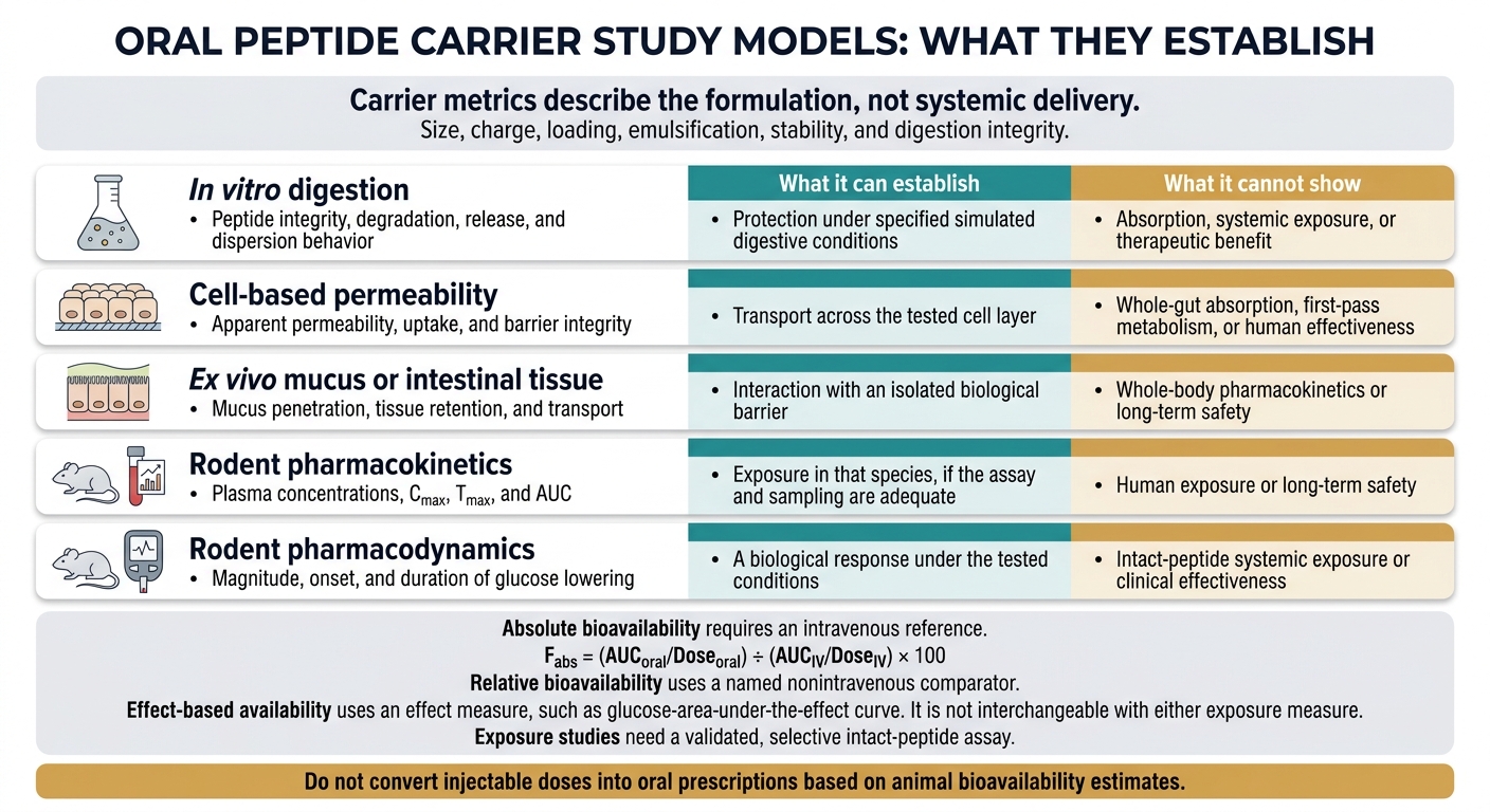
Oral Peptide Carriers: What Each Study Model Can Prove
What Each Study Model Can Show
The question isn’t whether a carrier looks promising. It’s which measured endpoint shows delivery.
Carrier metrics - size, charge, loading, emulsification, stability, and digestion integrity - describe the formulation, not systemic delivery. Digestion tests can show protection under simulated conditions. They don’t prove absorption.
| Study model | Measured endpoint | What it can establish | What it cannot show |
|---|---|---|---|
| In vitro digestion | Peptide integrity, degradation, release, and dispersion behavior | Protection under specified simulated digestive conditions | Absorption, systemic exposure, or therapeutic benefit |
| Cell-based permeability | Apparent permeability, uptake, and barrier integrity | Transport across the tested cell layer | Whole-gut absorption, first-pass metabolism, or human effectiveness |
| Ex vivo mucus or intestinal tissue | Mucus penetration, tissue retention, and transport | Interaction with an isolated biological barrier | Whole-body pharmacokinetics or long-term safety |
| Rodent pharmacokinetics | Plasma concentrations, Cmax, Tmax, and AUC | Exposure in that species, if the assay and sampling are adequate | Human exposure or long-term safety |
| Rodent pharmacodynamics | Magnitude, onset, and duration of glucose lowering | A biological response under the tested conditions | Intact-peptide systemic exposure or clinical effectiveness |
Bioavailability Calculations and Study Controls
Absolute bioavailability requires an intravenous reference:
Fabs = (AUCoral/Doseoral) ÷ (AUCIV/DoseIV) × 100.
Relative bioavailability uses a named nonintravenous comparator. Effect-based availability uses an effect measure, such as glucose-area-under-the-effect curve. It is not interchangeable with either exposure measure. Normalize doses to active peptide, and report species, route, and units.
Exposure studies need free-peptide, blank-carrier, and positive-comparator controls. They also need a validated, selective intact-peptide assay - not simply a measurement of “peptide-related” or immunoreactive material. Document the assay’s accuracy, precision, recovery, stability, and quantification limits.
Sampling should support AUC₀–t, AUC₀–∞, peak exposure, and terminal half-life. Look for sample-size justification, individual results, variability, confidence intervals, prespecified analyses and exclusions, and reported randomization or blinding.
Report the circulating analyte, not just the response. Longer activity may reflect delayed absorption or altered metabolism rather than sustained delivery. Test dose proportionality to help assess this distinction.
Limits of Animal Findings and Research Needs
Most published data are insulin-based, so these limits matter before extending claims to other peptides. Peptide classes differ in stability, potency, and receptor pharmacology. Translation to humans also depends on gastrointestinal pH, transit, mucus, enzymes, bile, fed state, and first-pass metabolism.
Before comparing results, record species, strain, sex, diabetic status, dose, administration volume, and feeding conditions. Inconsistent assays add another limit to cross-study comparisons. These gaps help shape the human pharmacokinetic/pharmacodynamic (PK/PD) studies needed next.
Stronger development programs need intravenous-referenced pharmacokinetics, intact-peptide assays, fed and fasted comparisons, and exposure-linked pharmacodynamic dose-ranging.
Repeat-dose toxicology should assess the complete formulation, including gastrointestinal tolerability, barrier function, liver and kidney findings, and excipient exposure. Ion-pairing agents and surfactants also need direct testing for tolerability and barrier effects.
Conclusion: What the Evidence Means for Clinical Practice
The preclinical data point to mechanisms that may work, not readiness for clinical use. Self-emulsifying carriers remain proof-of-concept systems, not evidence to support prescribing.
For insulin–sodium dodecyl sulfate SMEDDS, enteric-coated insulin SNEDDS, and insulin-glargine carriers, the reviewed evidence does not establish a validated oral dosing regimen in humans. Each finding applies only to the carrier, peptide, and model tested.
Do not convert injectable doses into oral prescriptions based on animal bioavailability estimates. Human exposure, efficacy, and safety data are still needed.
Prescriber Education and Regulatory Limits
For clinicians, regulatory status and human evidence matter more than whether a formulation is new.
PeptidePrescriber is an educational resource for licensed healthcare professionals. It does not provide evidence that these carriers work in humans.
FAQs
Could food affect oral peptide absorption?
Yes. Food can affect oral peptide absorption by delaying gastric emptying and changing lipid digestion. These changes may interfere with peptide release timing or destabilize self-emulsifying carriers.
Clinical studies should test delivery consistency under both fasted and standardized fed conditions. For BPC-157, the cited clinical guidance recommends taking it on an empty stomach to optimize delivery.
How can studies distinguish absorption from gut barrier damage?
Pair regional delivery measurements with safety and barrier-integrity assays. Track transepithelial electrical resistance (TEER) before, during, and after exposure. Also monitor tight-junction markers (ZO-1 and occludin) and FITC-dextran leakage. A lasting drop in resistance or marker leakage points to barrier damage.
Include mass balance checks and compare free peptide with peptide-loaded carriers. This helps separate carrier-specific effects from possible tissue damage caused by excipients.
Could these carriers work for peptides beyond insulin?
Yes. Researchers are studying self-emulsifying and other lipid-based carriers for peptides beyond insulin, though clinical use is still limited. These carriers aim to protect peptides from enzymatic breakdown, help them cross membranes, and prevent them from getting trapped in mucus.
Peptides such as KPV and BPC-157 have shown promise in preclinical research, but oral delivery remains experimental. Success depends on the peptide’s chemistry, the carrier’s design, and consistent bioavailability in humans - a requirement that still needs sufficient large-scale clinical validation.